. 2022 Sep 13;26(1):274.
doi: 10.1186/s13054-022-04159-x.

Systemic interleukin-6 inhibition ameliorates acute neuropsychiatric phenotypes in a murine model of acute lung injury

Affiliations

Systemic interleukin-6 inhibition ameliorates acute neuropsychiatric phenotypes in a murine model of acute lung injury

Faizan Anwar et al. Crit Care. .

Abstract

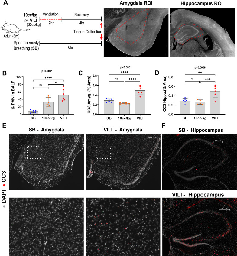
Acute neuropsychiatric impairments occur in over 70% of patients with acute lung injury. Mechanical ventilation is a well-known precipitant of acute lung injury and is strongly associated with the development of acute delirium and anxiety phenotypes. In prior studies, we demonstrated that IL-6 mediates neuropathological changes in the frontal cortex and hippocampus of animals with mechanical ventilation-induced brain injury; however, the effect of systemic IL-6 inhibition on structural and functional acute neuropsychiatric phenotypes is not known. We hypothesized that a murine model of mechanical ventilation-induced acute lung injury (VILI) would induce neural injury to the amygdala and hippocampus, brain regions that are implicated in diverse neuropsychiatric conditions, and corresponding delirium- and anxiety-like functional impairments. Furthermore, we hypothesized that these structural and functional changes would reverse with systemic IL-6 inhibition. VILI was induced using high tidal volume (35 cc/kg) mechanical ventilation. Cleaved caspase-3 (CC3) expression was quantified as a neural injury marker and found to be significantly increased in the VILI group compared to spontaneously breathing or anesthetized and mechanically ventilated mice with 10 cc/kg tidal volume. VILI mice treated with systemic IL-6 inhibition had significantly reduced amygdalar and hippocampal CC3 expression compared to saline-treated animals and demonstrated amelioration in acute neuropsychiatric behaviors in open field, elevated plus maze, and Y-maze tests. Overall, these data provide evidence of a pathogenic role of systemic IL-6 in mediating structural and functional acute neuropsychiatric symptoms in VILI and provide preclinical justification to assess IL-6 inhibition as a potential intervention to ameliorate acute neuropsychiatric phenotypes following VILI.

Keywords: Anxiety; Cleaved caspase-3; Delirium; IL-6; Neural injury; Neuroinflammation; VILI.


Conflict of interest statement

The authors declare that they have no competing interests.

Figures

Fig. 1
Fig. 1
Cleaved caspase-3 is significantly elevated in the amygdala and hippocampus of VILI mice. A Schematic of experimental design and representative regions of interest (ROIs) of measured brain regions. B Quantification of lung inflammation (%PMN in BALF). C–D Quantification of cleaved caspase-3 (CC3) percent area in the amygdala and the hippocampus where each dot represents one animal. E Representative amygdalar sections stained for CC3 (positive signal displayed in red) overlaid on DAPI nuclear stain (gray). Magnified regions of the indicated area of amygdala from the micrograph directly above. F Representative hippocampal sections stained for CC3 (positive signal displayed in red) overlaid on DAPI nuclear stain (gray). Quantitative data are expressed in mean ± SD. *p ≤ 0.05, **p ≤ 0.01, ***p ≤ 0.001, ****p ≤ 0.0001
Fig. 2
Fig. 2
VILI increases amygdalar neuronal activity, neuronal stress response, and both amygdalar and hippocampal IL-6 and TNF-α. A–B Quantified levels of cellular stress response (HSP90) and of neuronal activity (c-fos) within the amygdala and hippocampus. C–H IL-6 and TNF-α are significantly increased in VILI brains compared to SB controls in amygdala but not in the hippocampus, while there is no significant change in IL-1β in both areas. I Representative micrographs stained for IL-6 (positive signal displayed in red); cell nuclei are revealed by DAPI staining (gray). Quantitative data are expressed in mean ± SD. *p ≤ 0.05, **p ≤ 0.01, ***p ≤ 0.001, ****p ≤ 0.0001
Fig. 3
Fig. 3
Lung inflammation, cerebral IL-6, and cerebral TNF-α positively and significantly correlate with both amygdalar and hippocampal CC3. A–B Regression analysis between the percent of PMNs in bronchoalveolar lavage fluid (BALF) with amygdalar and hippocampal CC3, IL-6, or TNF-α. SB group is indicated in blue and VILI in red. Significance (p) and fitness (r2) values are indicated. C Regression analysis demonstrating significant positive relationships between amygdalar CC3 with IL-6 and TNF-α. D Micrographs of amygdalar cortical neurons stained for IL-6 (displayed in green) and CC3 (displayed in red). Magnified areas of individual neurons and glial cells are inset. Note that CC3-positive neurons also co-stain for IL-6 in VILI amygdala. E Regression analysis demonstrates a significant positive relationship between hippocampal CC3 with IL-6 but not with TNF-α
Fig. 4
Fig. 4
IL-6 inhibition significantly reduces amygdalar or hippocampal CC3 expression. A PMNs in BALF after VILI were significantly different from SB regardless of the three intervention groups (ANOVA, F = 9.861, dF = 21, p = 0.0005). There are no significant differences in oxygen saturations between the three VILI intervention groups; VILI + Saline, VILI + α-IL-6, and VILI + α-IL-6-receptor antibody. B–C Amygdalar (ANOVA, F = 8.738, dF = 21, p = 0.0009) and hippocampal (ANOVA, F = 9.806, dF = 21, p = 0.0005) CC3 expressions are significantly increased in the VILI + Saline group compared to SB control, but significantly reduced in both IL-6 inhibited groups. D–E IL-6 (ANOVA, F = 5.160, dF = 21, p = 0.0095) and TNF-α (ANOVA, F = 5.655, dF = 21, p = 0.0066) are significantly increased in the VILI + Saline group compared to SB control, but significantly reduced in both VILI + IL-6 inhibited groups. F There is no significant difference in percent area of amygdalar IL-1β between all groups. G–I Similar to amygdalar cytokines, IL-6 (ANOVA, F = 8.658, dF = 21, p = 0.0009) and TNF-α (ANOVA, F = 4.210, dF = 21, p = 0.0202) are significantly increased in the VILI + Saline group compared to SB control and significantly reduced in both VILI + IL-6 inhibiter groups; however, there is no difference in the percent area of IL-1β between all groups
Fig. 5
Fig. 5
Lung inflammation, cerebral IL-6, and cerebral TNF-α positively and significantly correlate with amygdalar and hippocampal CC3 after IL-6 inhibition. A Regression analysis between the percent of PMNs in bronchoalveolar lavage fluid with amygdalar CC3. SB group is indicated in blue and VILI + Saline in green, VILI + α-IL-6 in red, and VILI + α-IL-6-receptor in brown. Significance (p) and fitness (r2) values are indicated. B Regression analysis between the percent of PMNs in bronchoalveolar lavage fluid with hippocampal CC3. C–D Regression analysis shows a direct and significant relationship between IL-6 and CC3, and TNF-α and CC3 in amygdala and hippocampus
Fig. 6
Fig. 6
Systemic IL-6 inhibition significantly improves acute delirium- and anxiety-like neuropsychiatric functional impairments following VILI. A Schema of the apparatus and assessment of behavioral function in the center and peripheral zones of the Open Field. B–C In the open-field behavioral test, VILI + Saline mice spent significantly less time and traveled less distance in the center zone compared to the VILI + α-IL-6 group, indicating more anxiety behavior in the VILI + Saline group compared to the α-IL-6 treated group. D Schema of the elevated plus maze for behavioral assessment in open and closed arms. E–F In the elevated plus maze, the VILI + Saline mice entered and spent significantly less time in the open arms as compared to VILI + α-IL-6 mice, indicating increased anxiety and avoidance behaviors in the VILI + Saline group. G Schema of the Y-maze paradigm. H–I VILI + Saline mice had significantly less spontaneous alternations in the Y-maze compared to the α-IL-6 treated group, indicating impaired short-term memory. Quantitative data are expressed in mean ± SD. *p ≤ 0.05, **p ≤ 0.01, ***p ≤ 0.001, ****p ≤ 0.0001

References

    1. Sasannejad C, Ely EW, Lahiri S. Long-term cognitive impairment after acute respiratory distress syndrome: a review of clinical impact and pathophysiological mechanisms. Crit Care. 2019;23(1):352. doi: 10.1186/s13054-019-2626-z. - DOI - PMC - PubMed
    1. Hager DN, Dinglas VD, Subhas S, Rowden AM, Neufeld KJ, Bienvenu OJ, Touradji P, Colantuoni E, Reddy DR, Brower RG, et al. Reducing deep sedation and delirium in acute lung injury patients: a quality improvement project. Crit Care Med. 2013;41(6):1435–1442. doi: 10.1097/CCM.0b013e31827ca949. - DOI - PubMed
    1. Davydow DS, Desai SV, Needham DM, Bienvenu OJ. Psychiatric morbidity in survivors of the acute respiratory distress syndrome: a systematic review. Psychosom Med. 2008;70(4):512–519. doi: 10.1097/PSY.0b013e31816aa0dd. - DOI - PMC - PubMed
    1. Wu C, Chen X, Cai Y. Risk factors associated with acute respiratory distress syndrome and death in patients with coronavirus disease 2019 pneumonia in Wuhan, China (vol 180, pg 934, 2020) Jama Intern Med. 2020;180(7):1031–1031. doi: 10.1001/jamainternmed.2020.0994. - DOI - PMC - PubMed
    1. Girard TD, Shintani AK, Jackson JC, Gordon SM, Pun BT, Henderson MS, Dittus RS, Bernard GR, Ely EW. Risk factors for post-traumatic stress disorder symptoms following critical illness requiring mechanical ventilation: a prospective cohort study. Crit Care. 2007;11(1):R28. doi: 10.1186/cc5708. - DOI - PMC - PubMed