. 2022 Sep 13;19(1):149.
doi: 10.1186/s12985-022-01872-5.

Detection and discovery of plant viruses in soybean by metagenomic sequencing

Affiliations

Detection and discovery of plant viruses in soybean by metagenomic sequencing

Manjula G Elmore et al. Virol J. .

Abstract

Background: Viruses negatively impact soybean production by causing diseases that affect yield and seed quality. Newly emerging or re-emerging viruses can also threaten soybean production because current control measures may not be effective against them. Furthermore, detection and characterization of new plant viruses requires major efforts when no sequence or antibody-based resources are available.

Methods: In this study, soybean fields were scouted for virus-like disease symptoms during the 2016-2019 growing seasons. Total RNA was extracted from symptomatic soybean parts, cDNA libraries were prepared, and RNA sequencing was performed using high-throughput sequencing (HTS). A custom bioinformatic workflow was used to identify and assemble known and unknown virus genomes.

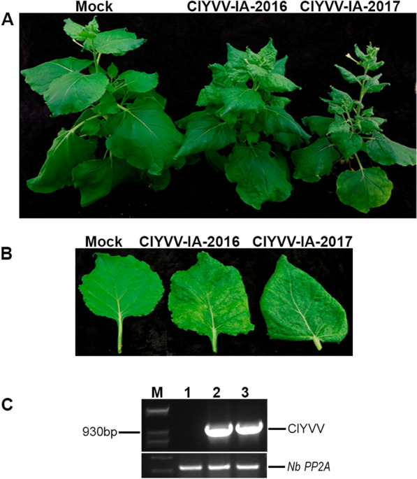
Results: Several viruses were identified in single or mixed infections. Full- or nearly full-length genomes were generated for tobacco streak virus (TSV), alfalfa mosaic virus (AMV), tobacco ringspot virus (TRSV), soybean dwarf virus (SbDV), bean pod mottle virus (BPMV), soybean vein necrosis virus (SVNV), clover yellow vein virus (ClYVV), and a novel virus named soybean ilarvirus 1 (SIlV1). Two distinct ClYVV isolates were recovered, and their biological properties were investigated in Nicotiana benthamiana, broad bean, and soybean. In addition to infections by individual viruses, we also found that mixed viral infections in various combinations were quite common.

Conclusions: Taken together, the results of this study showed that HTS-based technology is a valuable diagnostic tool for the identification of several viruses in field-grown soybean and can provide rapid information about expected viruses as well as viruses that were previously not detected in soybean.

Keywords: Broad bean; Clover yellow vein virus; High-throughput sequencing; Ilarvirus; Mixed infection; Nicotiana benthamiana; Soybean; Virus identification.


Conflict of interest statement

The authors declare that they have no competing interests.

Figures

Fig. 1
Fig. 1
Representative images of symptomatic soybean tissues collected from the field for metagenomics analysis. AH Leaves showing different patterns of chlorosis. A-H represent samples S1, S7, S12, S13, S18, S24, S25, and S26 respectively. I, J Examples of bud proliferation. I-J represent samples S4 and S14 respectively. K Stems thickened with brown corky necrosis. K represents sample S5. L, M Leaves exhibiting mottling. L-M represent samples S6 and S19 respectively. N, O Leaves with brown necrotic spots. N–O represent samples S39 and S40 respectively. P Leaves with angular yellow spots. P represents sample S41. QS Leaves displaying chlorosis of the veins or the tissue immediately surrounding the veins. Q-S represent samples S15, S16, and S17 respectively. T, U Leaves showing puckering and chlorosis. T-U represent samples S20 and S21 respectively. V, W Leaves exhibiting wilting and mosaic symptoms. V-W represent samples S22 and S23 respectively. X Pods with necrotic spots. X represent sample S24. Y, Z Leaves that rapidly became generally chlorotic and then necrotic. Y–Z represent samples S27, S28 respectively
Fig. 2
Fig. 2
Maximum likelihood phylogenetic tree showing the relationship of the ClYVV-IA isolates to other ClYVV isolates and BYMV. The phylogenetic tree was based on the amino acid sequences of ClYVV and Bean yellow mosaic virus (BYMV). BYMV is included as an outgroup. This analysis used the fully sequenced genomes available in GenBank, except for ClYVV isolate contig27.1 Ohio USA, which was a partial sequence. Sequences were aligned using MUSCLE, and the tree constructed from the alignment using PhyML. The tree branches were bootstrapped with 1000 replications. The de novo assembled ClYVV-IA genomes identified in this study, along with their respective accession number are denoted by the red text and (*) star
Fig. 3
Fig. 3
Symptoms of ClYVV-IA-2016 and ClYVV-IA-2017 in systemically infected leaves of N. benthamiana. Healthy mock-inoculated controls are indicated. Soybean field samples infected with ClYVV-IA-2016 or ClYVV-IA-2017 was used as an inoculum to infect N. benthamiana plants by mechanical inoculation. A Plants infected with ClYVV IA-2016 and ClYVV IA-2017 at 21 days post inoculation (dpi). B Systemic leaf with mosaic chlorosis and epinasty symptoms at 21 dpi. C RT-PCR detection of ClYVV isolates in N. benthamiana infected plants. Agarose gel electrophoresis of PCR confirming the presence of ClYVV-IA-2016 and ClYVV-IA-2017 identified by RNA sequencing. Lane 1: Mock- inoculated control; 2: Detection of ClYVV-IA-2016; 3: Detection of ClYVV-IA-2017; M: 1 Kb plus molecular weight maker. Sanger sequencing confirmed amplicon identities. N. benthamiana Protein phosphatase 2A (PP2A) reference gene was used as internal controls
Fig. 4
Fig. 4
Symptoms caused by ClYVV-IA-2016 and ClYVV-IA-2017 in broad bean. Healthy mock-inoculated controls are indicated. N. benthamiana infected with ClYVV-IA-2016 or ClYVV-IA-2017 was used as an inoculum to infect broad bean plants by mechanical inoculation. A Whole plants (cv. Broad Windsor) infected with ClYVV IA-2016 and ClYVV IA-2017 at 21 days post inoculation (dpi). B Systemic leaves (cv. Broad Windsor) showing symptoms at 21 dpi. C Whole plants (cv. Robin Hood) infected with ClYVV IA-2016 and ClYVV IA-2017 at 21 dpi. D Systemic leaves (cv. Robin Hood) showing symptoms at 21 dpi. (E) RT-PCR detection of ClYVV isolates in broad bean infected plants. Agarose gel electrophoresis of PCR confirming the presence of ClYVV-IA-2016 and ClYVV-IA-2017 identified by RNA sequencing. Lanes 1 and 4: Mock- inoculated controls of Broad Windsor and Robin Hood respectively; 2 and 5: Detection of ClYVV-IA-2016 in Broad Windsor and Robin Hood respectively; 3 and 6: Detection of ClYVV-IA-2017 in Broad Windsor and Robin Hood respectively; M: 1 Kb plus molecular weight maker. Sanger sequencing confirmed amplicon identities. Broad bean cyclophilin (CYP2) reference gene was used as internal controls
Fig. 5
Fig. 5
Symptoms caused by ClYVV-IA-2016 and ClYVV-IA-2017 in soybean. Healthy mock-inoculated controls are indicated. N. benthamiana infected with ClYVV-IA-2016 or ClYVV-IA-2017 was used as an inoculum to infect soybean plants by mechanical inoculation. A Whole plants infected with ClYVV IA-2016 at 35 days post inoculation (dpi). B Systemic leaf symptoms at 35 dpi. A representative young leaflet is shown in the middle of the panel while mature older leaflet is shown on the right. C Whole plants infected ClYVV IA-2017 at 21 dpi. D Systemic leaf symptoms at 21 dpi. A representative young trifoliolate leaf is shown in the middle of the panel while a mature older leaflet is shown on the right. E RT-PCR detection of ClYVV isolates in soybean infected plants. Agarose gel electrophoresis of PCR confirming the presence of ClYVV-IA-2016 and ClYVV-IA-2017 identified by RNA sequencing. Lane 1: Mock- inoculated control; 2: Detection of ClYVV-IA-2016; 3: Detection of ClYVV-IA-2017; M: 1 Kb plus molecular weight maker. Sanger sequencing confirmed amplicon identities. Soybean cyclophilin (CYP2) reference gene was used as internal controls
Fig. 6
Fig. 6
Transmission electron micrographs showing the ultrastructure of N. benthamiana mock-inoculated and ClYVV (IA-2017 isolate) infected leaf tissues A Mock inoculated leaf tissue at 10 dpi. Uninfected phloem parenchymal cell (PPC) surrounded by mesophyll cells (MC). Bar = 2 μm. (B-F) ClYVV infected leaf tissue at 10 dpi. B Infected vascular bundle surrounded by mesophyll cells (MC). Crystalline nuclear inclusions (NI) and cylindrical cytoplasmic inclusions (CI) distribution in the phloem cell parenchyma (PCP). Bar = 5 μm. C Infected mesophyll cells (MC). Crystalline nuclear inclusions (NI) and cylindrical cytoplasmic inclusions (CI) distribution in adjacent mesophyll cells. Bar = 2 μm. D Infected vascular parenchymal cell (VPC) above a xylem element (XE) and mesophyll cell (MC). Crystalline nuclear inclusions (NI) distribution in the nucleolus (Nu) and cytoplasm (Cy). Cylindrical cytoplasmic inclusions (CI) distribution in the cytoplasm. Bar = 2 μm. E Magnified virus particles from section of panel D. Virions (Vi), crystalline nuclear inclusions (NI) and cylindrical cytoplasmic inclusions (CI) distribution in the cytoplasm (Cy). Bar = 500 nm. F Magnified virus particles from section of panel D. Crystalline nuclear inclusions (NI) and cylindrical cytoplasmic inclusions (CI) distribution in the cytoplasm (Cy). Bar = 500 nm. C, chloroplast; St, starch; M, mitochondria; RER, rough endoplasmic reticulum; N, nucleus; NE, nuclear envelope; V, vacuole; CW, cell wall; PM, plasma membrane, PD, plasmodesmata; ICS, intracellular space; PC, phloem cell; Asterisk (*), phloem secondary cell wall thickening
Fig. 7
Fig. 7
Genome organization of soybean ilarvirus 1 (SIlV1). The line segments indicate the genome sizes, and the boxes indicate each open reading frame (ORF). The RNA methyltransferase (MET), RNA helicase (HEL), and viral RNA dependent RNA polymerase (RDRP) domains were identified using CD-search [67] and are indicated by black shading. The asterisk indicates that RNA2 is likely lacking a few bases at the 5’ end and is not full-length. Although the termini were not confirmed by rapid amplification of cDNA ends, the termini of RNA1 and RNA3 obtained from our sequence assembly are consistent with the full-length sequences of related viruses
Fig. 8
Fig. 8
Maximum likelihood phylogenetic tree showing the relationship of the soybean ilarvirus 1 (SIlV1) to other ilarviruses. The phylogenetic tree was based on the amino acid sequences of A 1a, B 2a, C MP, and D CP of SIlV1 and other ilarviruses. CMV is included as an outgroup. Sequences were aligned using MUSCLE, and the tree constructed from the alignment using PhyML. The tree branches were bootstrapped with 1000 replications. The de novo assembled SIlV1 genome identified in this study, along with its respective accession number are denoted by a red (*) star

References

    1. Wilson RF. Soybean: market driven research needs. In: Stacey G, editor. Genet genomics soybean. New York, NY: Springer; 2008. pp. 3–15.
    1. Ghabrial SA, Pickard CM, Stuckey RE. Identification and distribution of virus diseases of soybean in Kentucky. Plant Dis Rep. 1977;61:690–694.
    1. Harrison B, Steinlage TA, Domier LL, D’Arcy CJ. Incidence of Soybean dwarf virus and identification of potential vectors in Illinois. Plant Dis. 2005;89:28–32. doi: 10.1094/PD-89-0028. - DOI - PubMed
    1. Giesler LJ, Ziems AD. Incidence of Alfalfa mosaic virus, Bean pod mottle virus, and Soybean mosaic virus in Nebraska Soybean fields. Plant Health Prog. 2006;7:37. doi: 10.1094/PHP-2006-0424-01-HM. - DOI
    1. Bradshaw JD, Rice ME, Hill JH. Evaluation of management strategies for bean leaf beetles (Coleoptera: Chrysomelidae) and bean pod mottle virus (Comoviridae) in soybean. J Econ Entomol. 2008;101:1211–27. doi: 10.1093/jee/101.4.1211. - DOI - PubMed

Publication types

Supplementary concepts

LinkOut - more resources