Case Reports
. 2022 Feb 21;8(3):20210232.
doi: 10.1259/bjrcr.20210232. eCollection 2022 Sep.

Giant epiphrenic diverticulum: an unusual case from diagnosis to treatment

Affiliations
Case Reports

Giant epiphrenic diverticulum: an unusual case from diagnosis to treatment

Francesca Gorgoglione et al. BJR Case Rep. .

Abstract

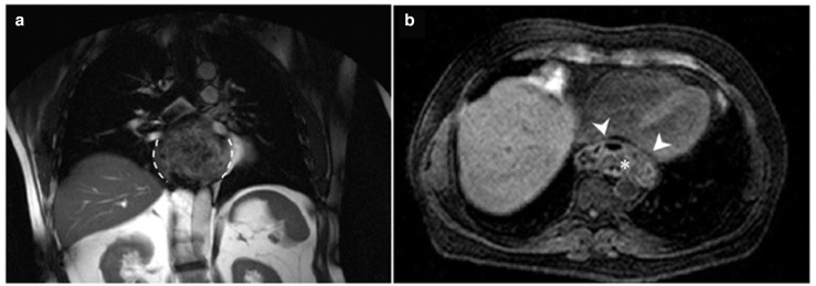
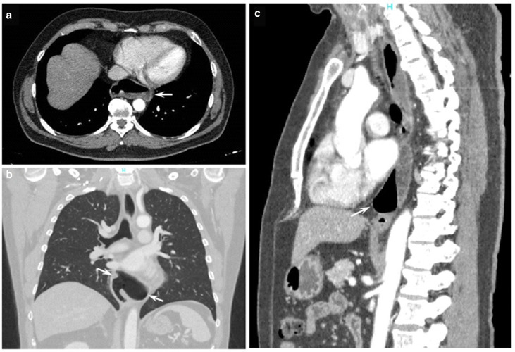
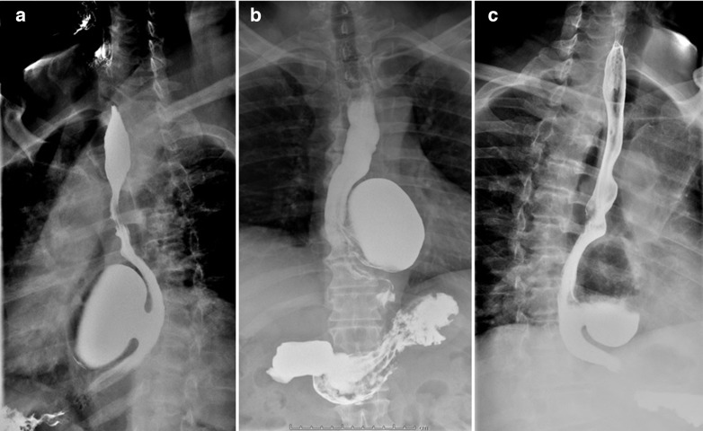
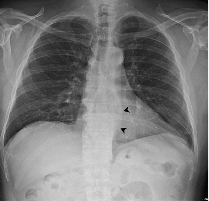
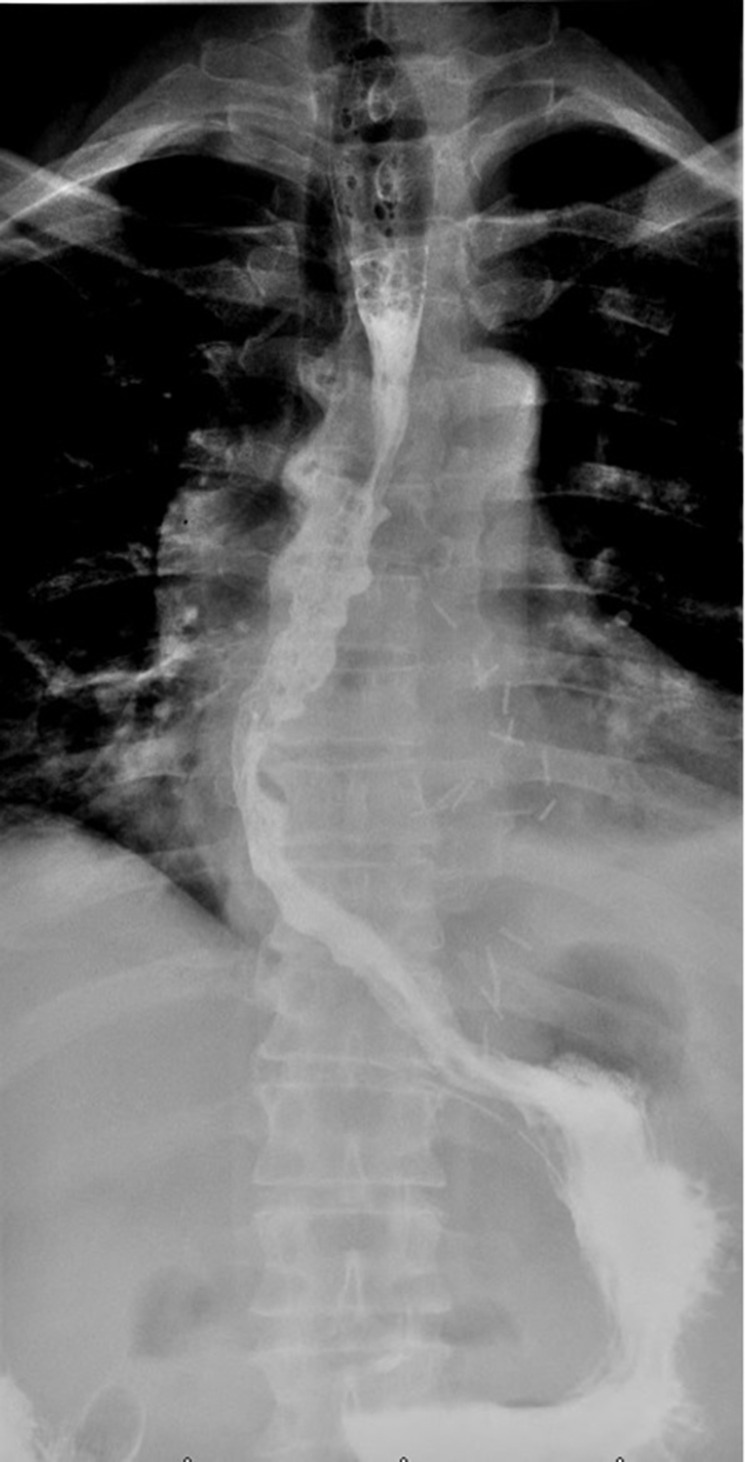
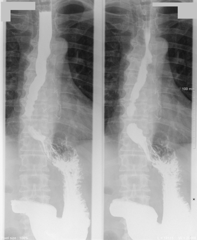
Esophageal diverticulum is a rare disease caused by impairment of esophageal motility. The incidence is not known, due to lack of symptoms in many cases. Surgical treatment is reserved to symptomatic patients. In this case report, we describe a rare case of epiphrenic esophageal diverticulum. A 61-year-old male with silent medical history, suffering severe chest pain had a CT scan showing a large esophageal diverticulum. The patient was referred to our hospital, IRCCS "Casa Sollievo della Sofferenza", to complete pre-operative assessment with a CT scan and a Barium swallowing radiography, giving morphodimensional details of the diverticulum. Based on these findings, the surgeons have chosen the appropriate operative strategy. The surgeons adopted a laparoscopic access, completed with robotic-assisted laparotomy due to the morphology of the diverticulum. Radiological evaluation is crucial in the diagnosis and in the treatment planning of symptomatic patients.


Figures

Figure 1.
Figure 1.
(A) Coronal T1W MRI demonstrated the left-sided large epiphrenic diverticulum of the esophagus (dashed lines); (B) axial thrive MRI showed the esophageal diverticulum compressing the left atrium and ventricle (arrowhead); it also evidenced the uneven content of the diverticulum, presumably food debris (asterisk).
Figure 2.
Figure 2.
Pre-operative CT scan: June 2021. (A) Axial CE CT chest image in soft tissue window demonstrated a large outpouching on the left wall in the lower esophagus (arrow), with air-fluid level, due to the large esophageal diverticulum; (B) coronal CE CT chest image in lung window clearly showed the large left-sided esophageal diverticulum communicating with the esophagus (arrow); (C) sagittal CE CT chest in soft tissue window image showed the retrocardiac extension of the diverticulum (arrow).
Figure 3.
Figure 3.
Barium swallowing X-ray showed the large epiphrenic esophageal diverticulum on the left anterior wall with 2,5 cm neck, in the right oblique projection (A), AP projection (B) and left oblique projection (C). AP, anteroposterior.
Figure 4.
Figure 4.
Pre-operative chest X-ray showing the wall of the diverticulum (arrowheads).
Figure 5.
Figure 5.
7 days post-surgery esophagogram with water soluble iodinated contrast medium showed no abnormal leaking of contrast.
Figure 6.
Figure 6.
3 months post-surgery, Barium swallowing X-ray showed no recurrence of diverticulum.

References

    1. Clark SC, Norton SA, Jeyasingham K, Ridley PD. Oesophageal epiphrenic diverticulum: an unusual presentation and review. Ann R Coll Surg Engl 1995; 77: 342–45. - PMC - PubMed
    1. Leandri C, Soyer P, Oudjit A, Guillaumot MA, Chaussade S, et al. . Contribution of magnetic resonance imaging to the management of esophageal diseases: a systematic review. Eur J Radiol 2019; 120: 108684. doi: 10.1016/j.ejrad.2019.108684 - DOI - PubMed
    1. Fisichella PM, Jalilvand A, Dobrowolsky A. Achalasia and epiphrenic diverticulum. World J Surg 2015; 39: 1614–19. doi: 10.1007/s00268-015-2950-7 - DOI - PubMed
    1. Hukkeri VS, Jindal S, Qaleem M, Tandon V, Govil D. Robotic transhiatal excision of epiphrenic diverticula. J Robot Surg 2016; 10: 365–68. doi: 10.1007/s11701-016-0595-7 - DOI - PubMed
    1. Bhandarwar AH, Tungenwar PN, Sawakare YM, Wagh AN, Patel CB, et al. . Large epiphrenic diverticula: a rare case presentation. Clin Pract 2015; 5: 784. doi: 10.4081/cp.2015.784 - DOI - PMC - PubMed

Publication types