. 2022 Sep 20;11(18):e026399.
doi: 10.1161/JAHA.122.026399. Epub 2022 Sep 14.

Assessment of Cardiac, Vascular, and Pulmonary Pathobiology In Vivo During Acute COVID-19

Affiliations

Assessment of Cardiac, Vascular, and Pulmonary Pathobiology In Vivo During Acute COVID-19

Shirjel R Alam et al. J Am Heart Assoc. .

Abstract

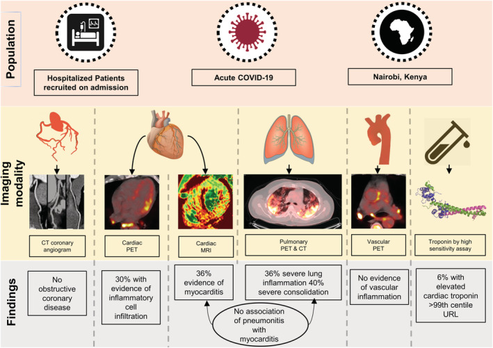
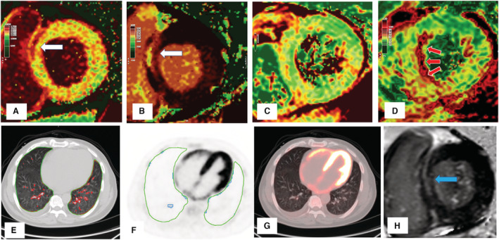
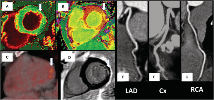
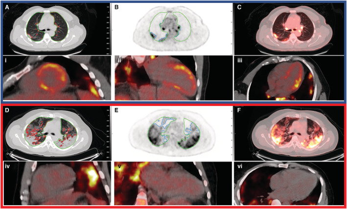
Background Acute COVID-19-related myocardial, pulmonary, and vascular pathology and how these relate to each other remain unclear. To our knowledge, no studies have used complementary imaging techniques, including molecular imaging, to elucidate this. We used multimodality imaging and biochemical sampling in vivo to identify the pathobiology of acute COVID-19. Specifically, we investigated the presence of myocardial inflammation and its association with coronary artery disease, systemic vasculitis, and pneumonitis. Methods and Results Consecutive patients presenting with acute COVID-19 were prospectively recruited during hospital admission in this cross-sectional study. Imaging involved computed tomography coronary angiography (identified coronary disease), cardiac 2-deoxy-2-[fluorine-18]fluoro-D-glucose positron emission tomography/computed tomography (identified vascular, cardiac, and pulmonary inflammatory cell infiltration), and cardiac magnetic resonance (identified myocardial disease) alongside biomarker sampling. Of 33 patients (median age 51 years, 94% men), 24 (73%) had respiratory symptoms, with the remainder having nonspecific viral symptoms. A total of 9 patients (35%, n=9/25) had cardiac magnetic resonance-defined myocarditis. Of these patients, 53% (n=5/8) had myocardial inflammatory cell infiltration. A total of 2 patients (5%) had elevated troponin levels. Cardiac troponin concentrations were not significantly higher in patients with and without myocarditis (8.4 ng/L [interquartile range, IQR: 4.0-55.3] versus 3.5 ng/L [IQR: 2.5-5.5]; P=0.07) or myocardial cell infiltration (4.4 ng/L [IQR: 3.4-8.3] versus 3.5 ng/L [IQR: 2.8-7.2]; P=0.89). No patients had obstructive coronary artery disease or vasculitis. Pulmonary inflammation and consolidation (percentage of total lung volume) was 17% (IQR: 5%-31%) and 11% (IQR: 7%-18%), respectively. Neither were associated with the presence of myocarditis. Conclusions Myocarditis was present in a third patients with acute COVID-19, and the majority had inflammatory cell infiltration. Pneumonitis was ubiquitous, but this inflammation was not associated with myocarditis. The mechanism of cardiac pathology is nonischemic and not attributable to a vasculitic process. Registration URL: https://www.isrctn.com; Unique identifier: ISRCTN12154994.

Keywords: CMR; COVID‐19; FDG‐PET; myocarditis; pneumonitis.


Figures

Figure 1
Figure 1. Study design.
Patients with acute COVID‐19 were scanned on hospital admission. Cardiac magnetic resonance revealed myocarditis in 1 in 3 patients using the most stringest diagnostic criteria. Myocardial inflammatory cell infiltration identified by 2‐deoxy‐2‐[fluorine‐18]fluoro‐D‐glucose PET/CT was present in 30% of all patients, and in the majority of patients with cardiac magnetic resonance–defined myocarditis. No patient had significant coronary artery disease on CT coronary angiography scanning. No patient had vasculitis. Although significant pulmonary inflammation and consolidation was common, it was not associated with the presence of myocarditis. Troponin testing did not identify patients with imaging evidence of myocardial edema or inflammatory cell infiltration. CT indicates computed tomography; MRI, magnetic resonance imaging; PET, positron emission tomography; and URL, upper reference limit.
Figure 2
Figure 2. Severe myocarditis with minimal lung injury.
There is mid‐wall injury at the basal myocardium in the septum (white arrows) shown by cardiac magnetic resonance (CMR) (A) native T1, (B) postcontrast T1, and late gadolinium enhancement (H; blue arrow). There is no increase in T2 values in this basal region (C), but there is gross increase in mid‐ventricular septal T2 (D; red arrows), indicating edema remote to prior myocardial fibrosis. There was minimal lung consolidation (E; red contours) or inflammation (F; blue contours). There is diffuse biventricular 2‐deoxy‐2‐[fluorine‐18]fluoro‐D‐glucose uptake (significantly higher than in the liver) (G). The patient had severe left and right ventricle impairment with elevated high‐sensitivity cardiac troponin I (110 ng/L) and NT‐proBNP (N‐terminal pro–brain natriuretic peptide; 7140 pg/mL) but low CRP (C‐reactive protein; 10 mg/L).
Figure 3
Figure 3. Focal inferolateral myocarditis with no atherosclerotic disease.
Changes (white arrows) in the native and postcontrast cardiac magnetic resonance (CMR) T1 values (A and B), 2‐deoxy‐2‐[fluorine‐18]fluoro‐D‐glucose positron emission tomography focal uptake (C), and subendocardial fibrosis on CMR late gadolinium enhancement (D). There was no significant coronary artery disease on computed tomography coronary angiography (E through G). Biochemical cardiac and inflammatory markers were low (high‐sensitivity cardiac troponin I, 2.72 ng/L; NT‐proBNP [N‐terminal pro–brain natriuretic peptide], <35 pg/mL; CRP [C‐reactive protein], 4 mg/L). Cx indicates left circumflex coronary artery; LAD, left anterior descending coronary artery; and RCA, right coronary artery.
Figure 4
Figure 4. Cardiac and pulmonary 2‐deoxy‐2‐[fluorine‐18]fluoro‐D‐glucose positron emission tomography/CT imaging in 2 patients showing discordance between pulmonary and myocardial involvement.
Top panel (blue outline) represents a patient with significant myocardial inflammatory cell infiltration with some pulmonary involvement—17% lung consolidation and 29% inflammation. Cardiac inflammatory cell infiltration (focal on diffuse bright spots in lateral anterior and septal walls). Bottom panel (red outline) represents another patient with no myocardial involvement but with significant lung consolidation (35%) and inflammation (54%). Lung consolidation on computed tomography (CT; A and D; red contours) and lung inflammation (B and E; blue contours) are shown. Green contours indicate lung parenchyma. Fused image (C and F) showing lung inflammation with heat maps on CT. Cardiac 2‐deoxy‐2‐[fluorine‐18]fluoro‐D‐glucose positron emission tomography shows inflammatory cell infiltration in the short axis (i and iv), 2‐chamber (ii and v), and 4‐chamber views (iii and vi).

References

    1. Puntmann VO, Carerj ML, Wieters I, Fahim M, Arendt C, Hoffmann J, Shchendrygina A, Escher F, Vasa‐Nicotera M, Zeiher AM, et al. Outcomes of cardiovascular magnetic resonance imaging in patients recently recovered from coronavirus disease 2019 (COVID‐19). JAMA Cardiol. 2020;5:1265–1273. doi: 10.1001/jamacardio.2020.3557 - DOI - PMC - PubMed
    1. Wang H, Li R, Zhou Z, Jiang H, Yan Z, Tao X, Li H, Xu L. Cardiac involvement in COVID‐19 patients: mid‐term follow up by cardiovascular magnetic resonance. J Cardiovasc Magn Reson. 2021;23:14. doi: 10.1186/s12968-021-00710-x - DOI - PMC - PubMed
    1. Kotecha T, Knight DS, Razvi Y, Kumar K, Vimalesvaran K, Thornton G, Patel R, Chacko L, Brown JT, Coyle C, et al. Patterns of myocardial injury in recovered troponin‐positive COVID‐19 patients assessed by cardiovascular magnetic resonance. Eur Heart J. 2021;42:1866–1878. doi: 10.1093/eurheartj/ehab075 - DOI - PMC - PubMed
    1. Zhou F, Yu T, Du R, Fan G, Liu Y, Liu Z, Xiang J, Wang Y, Song B, Gu X, et al. Clinical course and risk factors for mortality of adult inpatients with COVID‐19 in Wuhan, China: a retrospective cohort study. Lancet. 2020;395:1054–1062. doi: 10.1016/S0140-6736(20)30566-3 - DOI - PMC - PubMed
    1. Nensa F, Kloth J, Tezgah E, Poeppel TD, Heusch P, Goebel J, Nassenstein K, Schlosser T. Feasibility of FDG‐PET in myocarditis: comparison to CMR using integrated PET/MRI. J Nucl Cardiol. 2018;25:785–794. doi: 10.1007/s12350-016-0616-y - DOI - PubMed

Publication types

Associated data