

Body Surface Potential Mapping during Ventricular Depolarization in Rats after Acute Exhaustive Exercise
- PMID: 36102423
- PMCID: PMC9750213
- DOI: 10.36660/abc.20211058
Body Surface Potential Mapping during Ventricular Depolarization in Rats after Acute Exhaustive Exercise
Abstract
Background: Exhaustive physical exercise can cause substantial changes in the electrical properties of the myocardium.
Objective: To evaluate, using body surface potential mapping, the electrical activity of the heart in rats during ventricular depolarization after acute exhaustive exercise.
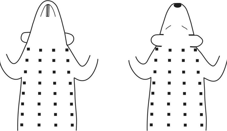
Methods: Twelve-week-old male rats were submitted to acute treadmill exercise at 36 m/min until exhaustion. Unipolar electrocardiograms (ECGs) from the torso surface were recorded in zoletil-anesthetized rats three to five days before (Pre-Ex), 5 and 10 minutes after exhaustive exercise (Post-Ex 5 and Post-Ex 10, respectively) simultaneously with ECGs in limb leads. The instantaneous body surface potential maps (BSPMs) were analyzed during ventricular depolarization. P values <0.05 were considered statistically significant.
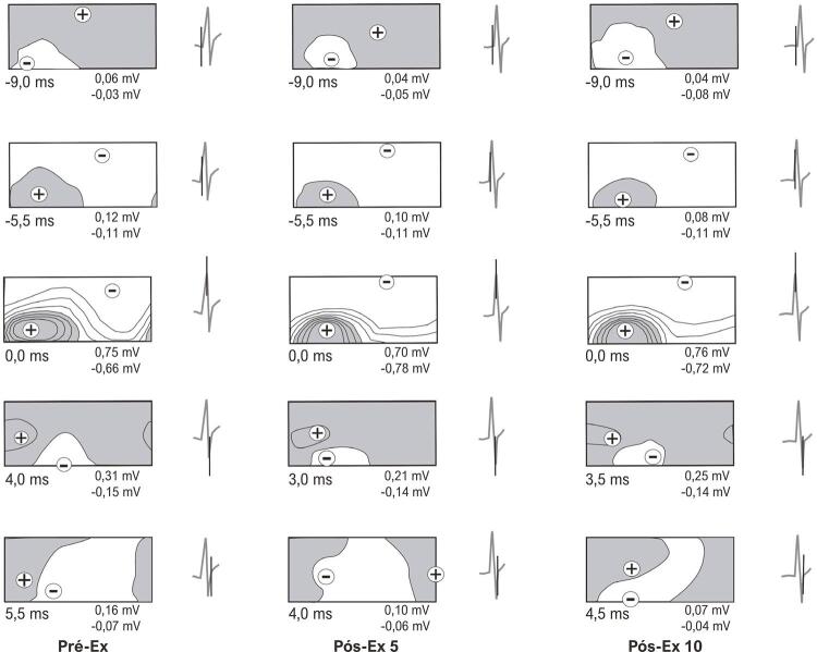
Results: Compared with Pre-Ex, an early completion of the second inversion of potential distributions, an early completion of ventricular depolarization, as well as a decrease in the duration of the middle phase and the total duration of ventricular depolarization on BSPMs were revealed at Post-Ex 5. Also, compared with Pre-Ex, an increase in the amplitude of negative BSPM extremum at the R-wave peak on the ECG in lead II (RII-peak) and a decrease in the amplitude of negative BSPM extremum at 3 and 4 ms after RII-peak were showed at Post-Ex 5. At Post-Ex 10, parameters of BSPMs did not differ from those at Pre-Ex.
Conclusion: In rats, acute exhaustive exercise causes reversible changes in the temporal and amplitude characteristics of BSPMs during ventricular depolarization, most likely related to alterations in the excitation of the main mass of the ventricular myocardium.
Fundamento: O exercício físico exaustivo pode causar alterações significantes nas propriedades elétricas do miocárdio.
Objetivo: Avaliar, através do mapeamento potencial de superfície corporal, a atividade elétrica do coração de ratos durante a despolarização ventricular após exercício exaustivo agudo.
Métodos: Ratos machos com doze semanas de idade foram submetidos a exercício agudo em esteira a 36 m/min até a exaustão. Eletrocardiogramas unipolares (ECGs) da superfície do tronco foram registrados em ratos anestesiados com zoletil três a cinco dias antes (Pré-Ex), 5 e 10 minutos após exercício exaustivo (Pós-Ex 5 e Pós-Ex 10, respectivamente) simultaneamente com ECGs nas derivações dos membros. Os mapas potenciais de superfície corporal instantâneos (BSPMs, body surface potential maps ) foram analisados durante a despolarização ventricular. Os valores de p <0,05 foram considerados estatisticamente significantes.
Resultados: Comparado com o Pré-Ex, uma conclusão precoce da segunda inversão de distribuições de potencial, uma conclusão precoce da despolarização ventricular, bem como uma diminuição na duração da fase média e a duração total da despolarização ventricular nos BSPMs foram reveladas no Pós-Ex5. Além disso, em comparação com o Pré-Ex, um aumento na amplitude do extremo negativo do BSPM no pico da onda R no ECG na derivação II (pico RII) e uma diminuição na amplitude do extremo negativo do BSPM a 3 e 4 ms após o pico RII foram demonstrados no Pós-Ex 5. No Pós-Ex 10, os parâmetros dos BSPMs não diferiram daqueles do Pré-Ex.
Conclusão: Em ratos, o exercício exaustivo agudo causa alterações reversíveis nas características temporais e de amplitude dos BSPMs durante a despolarização ventricular, provavelmente relacionadas a alterações na excitação da massa principal do miocárdio ventricular.
Conflict of interest statement
Potencial conflito de interesse
Não há conflito com o presente artigo.
Figures


Comment in
-
Applicability of Body Surface Potential Mapping Through Exercise in Small Animals.Arq Bras Cardiol. 2022 Nov;119(5):776-777. doi: 10.36660/abc.20220646. Arq Bras Cardiol. 2022. PMID: 36453769 Free PMC article. English, Portuguese. No abstract available.
References
LinkOut - more resources
Full Text Sources
