

Calcium and activity-dependent signaling in the developing cerebral cortex
- PMID: 36102617
- PMCID: PMC9578689
- DOI: 10.1242/dev.198853
Calcium and activity-dependent signaling in the developing cerebral cortex
Abstract
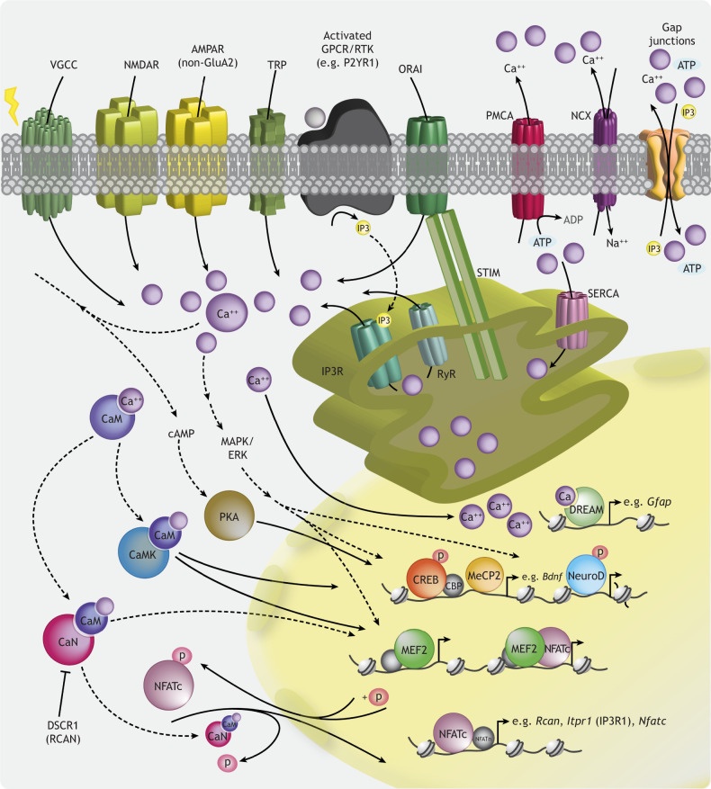
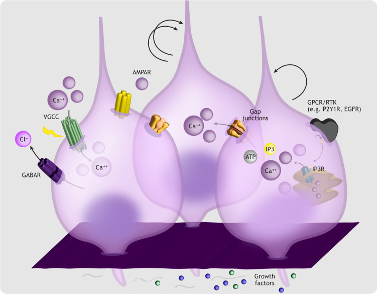
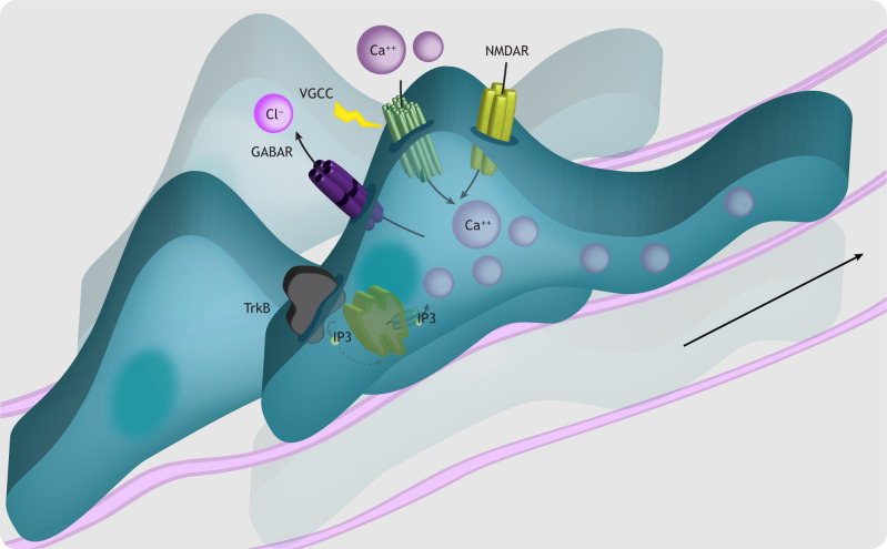
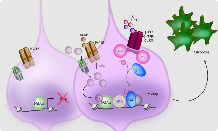
Calcium influx can be stimulated by various intra- and extracellular signals to set coordinated gene expression programs into motion. As such, the precise regulation of intracellular calcium represents a nexus between environmental cues and intrinsic genetic programs. Mounting genetic evidence points to a role for the deregulation of intracellular calcium signaling in neuropsychiatric disorders of developmental origin. These findings have prompted renewed enthusiasm for understanding the roles of calcium during normal and dysfunctional prenatal development. In this Review, we describe the fundamental mechanisms through which calcium is spatiotemporally regulated and directs early neurodevelopmental events. We also discuss unanswered questions about intracellular calcium regulation during the emergence of neurodevelopmental disease, and provide evidence that disruption of cell-specific calcium homeostasis and/or redeployment of developmental calcium signaling mechanisms may contribute to adult neurological disorders. We propose that understanding the normal developmental events that build the nervous system will rely on gaining insights into cell type-specific calcium signaling mechanisms. Such an understanding will enable therapeutic strategies targeting calcium-dependent mechanisms to mitigate disease.
Keywords: Calcium signaling; Cortical development; Neurodevelopmental disorders.
© 2022. Published by The Company of Biologists Ltd.
Conflict of interest statement
Competing interests The authors declare no competing or financial interests.
Figures
Similar articles
-
A Critical Neurodevelopmental Role for L-Type Voltage-Gated Calcium Channels in Neurite Extension and Radial Migration.J Neurosci. 2018 Jun 13;38(24):5551-5566. doi: 10.1523/JNEUROSCI.2357-17.2018. Epub 2018 May 17. J Neurosci. 2018. PMID: 29773754 Free PMC article.
-
Cadmium-induced apoptosis in primary rat cerebral cortical neurons culture is mediated by a calcium signaling pathway.PLoS One. 2013 May 31;8(5):e64330. doi: 10.1371/journal.pone.0064330. Print 2013. PLoS One. 2013. PMID: 23741317 Free PMC article.
-
Vasopressin-induced calcium signaling in cultured cortical neurons.Brain Res. 1998 May 18;793(1-2):244-54. doi: 10.1016/s0006-8993(98)00185-1. Brain Res. 1998. PMID: 9630655
-
Mapping the molecular and cellular complexity of cortical malformations.Science. 2021 Jan 22;371(6527):eaba4517. doi: 10.1126/science.aba4517. Science. 2021. PMID: 33479124 Review.
-
Exploring the intricacies of calcium dysregulation in ischemic stroke: Insights into neuronal cell death and therapeutic strategies.Life Sci. 2024 Jun 15;347:122651. doi: 10.1016/j.lfs.2024.122651. Epub 2024 Apr 19. Life Sci. 2024. PMID: 38642844 Review.
Cited by
-
Opto-chemogenetic inhibition of L-type CaV1 channels in neurons through a membrane-assisted molecular linkage.Cell Rep Methods. 2024 Nov 18;4(11):100898. doi: 10.1016/j.crmeth.2024.100898. Epub 2024 Nov 7. Cell Rep Methods. 2024. PMID: 39515337 Free PMC article.
-
Pulsed electromagnetic stimulation promotes neuronal maturation by up-regulating cholesterol biosynthesis.Stem Cell Res Ther. 2025 Jul 26;16(1):406. doi: 10.1186/s13287-025-04469-1. Stem Cell Res Ther. 2025. PMID: 40713855 Free PMC article.
-
Distinct calcium sources regulate temporal profiles of NMDAR and mGluR-mediated protein synthesis.Life Sci Alliance. 2024 May 15;7(8):e202402594. doi: 10.26508/lsa.202402594. Print 2024 Aug. Life Sci Alliance. 2024. PMID: 38749544 Free PMC article.
-
A 3D human iPSC-derived multi-cell type neurosphere system to model cellular responses to chronic amyloidosis.J Neuroinflammation. 2025 Apr 24;22(1):119. doi: 10.1186/s12974-025-03433-3. J Neuroinflammation. 2025. PMID: 40275379 Free PMC article.
-
ADNP dysregulates methylation and mitochondrial gene expression in the cerebellum of a Helsmoortel-Van der Aa syndrome autopsy case.Acta Neuropathol Commun. 2024 Apr 18;12(1):62. doi: 10.1186/s40478-024-01743-w. Acta Neuropathol Commun. 2024. PMID: 38637827 Free PMC article.
References
-
- Aguado, F., Carmona, M. A., Pozas, E., Aguiló, A., Martínez-Guijarro, F. J., Alcantara, S., Borrell, V., Yuste, R., Ibañez, C. F. and Soriano, E. (2003). BDNF regulates spontaneous correlated activity at early developmental stages by increasing synaptogenesis and expression of the K+/Cl- co-transporter KCC2. Development 130, 1267-1280. 10.1242/dev.00351 - DOI - PubMed
-
- Allen, M., Huang, B. S., Notaras, M. J., Lodhi, A., Barrio-Alonso, E., Lituma, P. J., Wolujewicz, P., Witztum, J., Longo, F., Chen, M.et al. (2022). Astrocytes derived from ASD individuals alter behavior and destabilize neuronal activity through aberrant Ca2+ signaling. Mol. Psychiatry 27, 2470-2484. 10.1038/s41380-022-01486-x - DOI - PMC - PubMed
Publication types
MeSH terms
Substances
Grants and funding
LinkOut - more resources
Full Text Sources
Medical
Research Materials
