. 2022 Sep 15;135(18):jcs260244.
doi: 10.1242/jcs.260244. Epub 2022 Sep 14.

Advances on the roles of tenascin-C in cancer

Affiliations

Advances on the roles of tenascin-C in cancer

Alev Yilmaz et al. J Cell Sci. .

Abstract

The roles of the extracellular matrix molecule tenascin-C (TNC) in health and disease have been extensively reviewed since its discovery over 40 years ago. Here, we will describe recent insights into the roles of TNC in tumorigenesis, angiogenesis, immunity and metastasis. In addition to high levels of expression in tumors, and during chronic inflammation, and bacterial and viral infection, TNC is also expressed in lymphoid organs. This supports potential roles for TNC in immunity control. Advances using murine models with engineered TNC levels were instrumental in the discovery of important functions of TNC as a danger-associated molecular pattern (DAMP) molecule in tissue repair and revealed multiple TNC actions in tumor progression. TNC acts through distinct mechanisms on many different cell types with immune cells coming into focus as important targets of TNC in cancer. We will describe how this knowledge could be exploited for cancer disease management, in particular for immune (checkpoint) therapies.

Keywords: Immunity; Metastasis; Tenascin-C; Tumor.


Conflict of interest statement

Competing interests G.O. has filed patents on the discovery of TNC targeting peptides (WO2021233766A1) and nanobodies (EP21305210.3). All other authors declare no competing or financial interests.

Figures

Fig. 1.
Fig. 1.
Chromosomal locus encoding TNC and ET-20, and domain structure and interaction partners of TNC. (A) Chromosomal location of the TNC protein-encoding sequence and sequence for the ET-20 lncRNA. The human TNC gene is shown schematically, with coding exons as dark blue boxes and non-coding exons as white boxes outlined with blue. The two exons in light blue encode the variable FNIII domains AD2 and AD1; these are not part of ENST00000350763 but have been added here. The TNC gene is found on the reverse strand of Chromosome 9 between 115,120 K and 115,020 K. Variants of the lncRNA ET-20 are found on the forward strand. Sequences homologous to the first exon of the three murine ET-20 variants are found between TNC exons 19 (encoding FNIII D) and 20 (encoding part of FNIII 6). Similarly, homologous sequences for the remainder of the short ET-20s sequence partially overlap with TNC exon 19. The positions of the exons encoding the middle-sized ET-20m and long ET-20l are mapped here based on their locations in the mouse genome. ET-20l exon 5 is predicted to be found near 115,250 K. ET-20l exons 5–7 are present in the region that is depicted as a gap. (B) Cartoon of the human TNC protein. Interaction partners include ECM molecules (red), cell surface receptors (green), soluble molecules (blue) and others (black). Therapeutic molecules with known interaction sites, such as antibodies [BC-2, BC-4 and Teleukin (also known as F16)], nanobodies Nb3, Nb4 and MAREMO peptides are indicated (orange). Note, that the TN3–TN5 and FBG domains are hotspots for molecular interactions. In addition to TGFβ, CXCL12, CCL21 and TRAIL, other soluble factors also bind to the same domains (TN4 and/or TN5), and several integrins bind to the TN3 domain. Other molecules such as annexin II, contactin, CCN2, collagen V, fibrillin-2, von Willenbrand factor (vWF), periostin, MMP2 and MMP3, as well as streptococcus and HIV have also been found to bind TNC, but without information on where exactly.
Fig. 2.
Fig. 2.
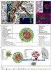
Illustration of TNC expression and function in tumors and lung metastasis. (A) Composite representation of TNC expression and function in the primary tumor and lung metastasis. Shown here are images derived from different tissues that have been assembled to provide a comprehensive visual display of the different sites of TNC expression. (i) Scanning electron micrograph (SEM) image of a PNET model tumor overexpressing TNC showing irregular and dysfunctional blood vessels with a small lumen diameter. Reproduced from Saupe et al. (2013), where it was published under a CC BY-NC-ND 3.0 license, under rights retained by G.O. (ii–v) Images of primary tumors with tissue staining for TNC in either green (immunofluorescence staining; v), red (immunofluorescence staining; iv) or brown [immunohistochemical (IHC) staining; iii]. (ii) Endothelial cells (CD31, red) are surrounded by freshly deposited TNC (arrows), reminiscent of pro-angiogenic TNC properties. Shown here is a human colorectal carcinoma. Reproduced from Spenlé et al. (2015a), where it was published under a CC-BY-NC-3.0 license, under rights retained by G.O. (iii) Vessel pruning. Continuum of an intact blood vessel (red blood cells inside lumen, asterisk) and pruned blood vessel (arrows) as determined by IHC indicating anti-angiogenic TNC properties. Shown here is a human insulinoma. Reproduced from Spenlé et al. (2015a), where it was published under a CC-BY-NC-3.0 license, under rights retained by G.O. (iv) TNC is deposited in several parallel aligned matrix fibers that generate matrix tracks (brown). The insert shows an enlarged image of such a track stained for laminin (LM, green) flanking TNC (red). Shown here is a murine PNET tumor. Reproduced from Spenlé et al. (2015a), where it was published under a CC-BY-NC-3.0 license, under rights retained by G.O. (v) Matrix tracks stained for TNC (green) and LM (white) separate tumor nests (DAPI, blue) and retained leukocytes (CD11c, red) inside the stroma. Shown here is a 4NQO-induced tongue OSCC. Reproduced from Spenlé et al. (2020) under rights retained by G.O. Fig. 2A is not published under the terms of the CC-BY license of this article. For permission to reuse, please see individual references. (B) TNC expression in lung metastasis. (i,ii) Pulmonary blood vessel with BVI as determined by either H&E (i) or staining for TNC (green) (ii). Clusters of CTC (CK8/18, red) are surrounded by TNC (green) and an outer endothelial monolayer. (iii) TNC expression in fibrillar matrix inside a pulmonary metastasis is reminiscent of matrix tracks seen in the primary tumor. Shown here is a MMTV-NeuNT tumor. Reproduced from Sun et al. (2019), where it was published under a CC-BY 4.0 license. Scale bars: 20 µm. (C) Regulation of cell phenotypes by TNC. Many tumors are compartmentalized into tumor cell nests that are demarcated by the surrounding stroma, which is inherently rich in TNC (green). TNC regulates the phenotypes of tumor cells, fibroblasts, ECs and immune cells as indicated, altogether orchestrating an immunosuppressive tumor microenvironment and promoting metastasis. BVI, blood vessel invasion; EMT, epithelial to mesenchymal transition; EV, extracellular vesicle; TAM, tumor-associated macrophage; TDEC, tumor cell-derived EC; TIL, tumor infiltrating leukocyte; Treg, T regulatory cells. (D) Effects of TNC-regulated signaling on the formation of local matrix niches in the primary tumor and at the metastatic site. Some molecules and pathways, discussed in this and other reviews, have been described to be exploited by TNC in the tumor and the metastatic niche. (i) Tumor niche. Schematic illustration of a tumor–stroma unit. Although DCs and tumor cells are expressing TNC, fibroblasts are the major source of TNC. Tumor cells and fibroblasts also express soluble factors that bind to TNC, which attract and retain TILs (DC, macrophages and CD8T cells) in the stroma. TNC also promotes infiltration of Treg into the tumor nest. (ii) Metastatic niche. Tumor cells and fibroblasts secrete EVs that travel through the circulation, either depositing TNC or inducing its expression at the metastatic site in the lung or lymph node. In addition, CTCs can also leave the primary tumor upon EMT or via amoeboid migration and reach the metastatic site, potentially attracted by TNC and/or TNC-induced factors. TNC promotes cell survival in CTCs while they circulate in the blood stream. EMT and the EC layers from the BVI promote pulmonary tissue entry. Crosstalk between tumor cells, macrophages and EC is instrumental in formation of the metastatic niche.
Fig. 3.
Fig. 3.
TNC regulates tumor immunity. (A) TNC can have opposing effects on tumor immunity. TNC might act as a DAMP by upregulating genes involved in antigen processing and presentation, but it also promotes Treg infiltration, upregulation of immune checkpoint molecules and the formation of tumor matrix tracks (TMTs) that immobilize a subset of TILs in the stroma. (B) As soon as TNC is expressed, TILs might be attracted into the tumor by factors expressed by the tumor cells (such as CXCL12), thus supporting immune surveillance (top). However, as soon as TMTs are formed, the immune surveillance and DAMP function of TNC is overcome by TIL retention, thus blocking access to the tumor cells (bottom). (C) Representative images of dendritic cells (CD11c, red) (i), and Treg (FoxP3, red) (ii) from 4NQO tongue OSCC. Reproduced from Spenlé et al. (2020) under rights retained by G.O. Fig. 3C is not published under the terms of the CC-BY license of this article. For permission to reuse, please see individual reference. Scale bars: 50 µm. The white arrows point at TILs retained in the stroma (I) or attracted into the tumor nest (II).
Fig. 4.
Fig. 4.
Targeting the immune functions of tenascin-C in cancer. (A) Summarized here are approaches for targeting the diverse functions of TNC in immunity that could be used to improve the anti-tumor immune surveillance phenotype. (B) Targeting ‘TIL-matrix retention’ represents a novel opportunity to target TNC in cancer, potentially abolishing the immune-exclusion phenotype and empowering immune checkpoint therapies that do not require the lowering of TNC expression, offering the opportunity to potentially preserve its desirable DAMP actions.

Similar articles

Cited by

References

    1. Albacete-Albacete, L., Navarro-Lérida, I., López, J. A., Martín-Padura, I., Astudillo, A. M., Ferrarini, A., Van-Der-Heyden, M., Balsinde, J., Orend, G., Vázquez, J.et al. (2020). ECM deposition is driven by caveolin-1–dependent regulation of exosomal biogenesis and cargo sorting. J. Cell Biol. 219, e202006178. 10.1083/jcb.202006178 - DOI - PMC - PubMed
    1. Angel, I., Pilo Kerman, O., Rousso-Noori, L. and Friedmann-Morvinski, D. (2020). Tenascin C promotes cancer cell plasticity in mesenchymal glioblastoma. Oncogene 39, 6990-7004. 10.1038/s41388-020-01506-6 - DOI - PubMed
    1. Asparuhova, M. B., Ferralli, J., Chiquet, M. and Chiquet-Ehrismann, R. (2011). The transcriptional regulator megakaryoblastic leukemia–1 mediates serum response factor–independent activation of tenascin–C transcription by mechanical stress. FASEB J. 25, 3477-3488. 10.1096/fj.11-187310 - DOI - PubMed
    1. Aubert, A., Mercier-Gouy, P., Aguero, S., Berthier, L., Liot, S., Prigent, L., Alcaraz, L. B., Verrier, B., Terreux, R., Moali, C.et al. (2021). Latent TGF-β activation is a hallmark of the tenascin family. Front. Immunol. 12, 613438. 10.3389/fimmu.2021.613438 - DOI - PMC - PubMed
    1. Bhattacharyya, S., Midwood, K. S. and Varga, J. (2022). Tenascin-C in fibrosis in multiple organs: translational implications. Semin. Cell Dev. Biol. 128, 130-136. 10.1016/j.semcdb.2022.03.019 - DOI - PMC - PubMed

Publication types