

Additive value of [18F]PI-2620 perfusion imaging in progressive supranuclear palsy and corticobasal syndrome
- PMID: 36102964
- PMCID: PMC9816230
- DOI: 10.1007/s00259-022-05964-w
Additive value of [18F]PI-2620 perfusion imaging in progressive supranuclear palsy and corticobasal syndrome
Abstract
Purpose: Early after [18F]PI-2620 PET tracer administration, perfusion imaging has potential for regional assessment of neuronal injury in neurodegenerative diseases. This is while standard late-phase [18F]PI-2620 tau-PET is able to discriminate the 4-repeat tauopathies progressive supranuclear palsy and corticobasal syndrome (4RTs) from disease controls and healthy controls. Here, we investigated whether early-phase [18F]PI-2620 PET has an additive value for biomarker based evaluation of 4RTs.
Methods: Seventy-eight patients with 4RTs (71 ± 7 years, 39 female), 79 patients with other neurodegenerative diseases (67 ± 12 years, 35 female) and twelve age-matched controls (69 ± 8 years, 8 female) underwent dynamic (0-60 min) [18F]PI-2620 PET imaging. Regional perfusion (0.5-2.5 min p.i.) and tau load (20-40 min p.i.) were measured in 246 predefined brain regions [standardized-uptake-value ratios (SUVr), cerebellar reference]. Regional SUVr were compared between 4RTs and controls by an ANOVA including false-discovery-rate (FDR, p < 0.01) correction. Hypoperfusion in resulting 4RT target regions was evaluated at the patient level in all patients (mean value - 2SD threshold). Additionally, perfusion and tau pattern expression levels were explored regarding their potential discriminatory value of 4RTs against other neurodegenerative disorders, including validation in an independent external dataset (n = 37), and correlated with clinical severity in 4RTs (PSP rating scale, MoCA, activities of daily living).
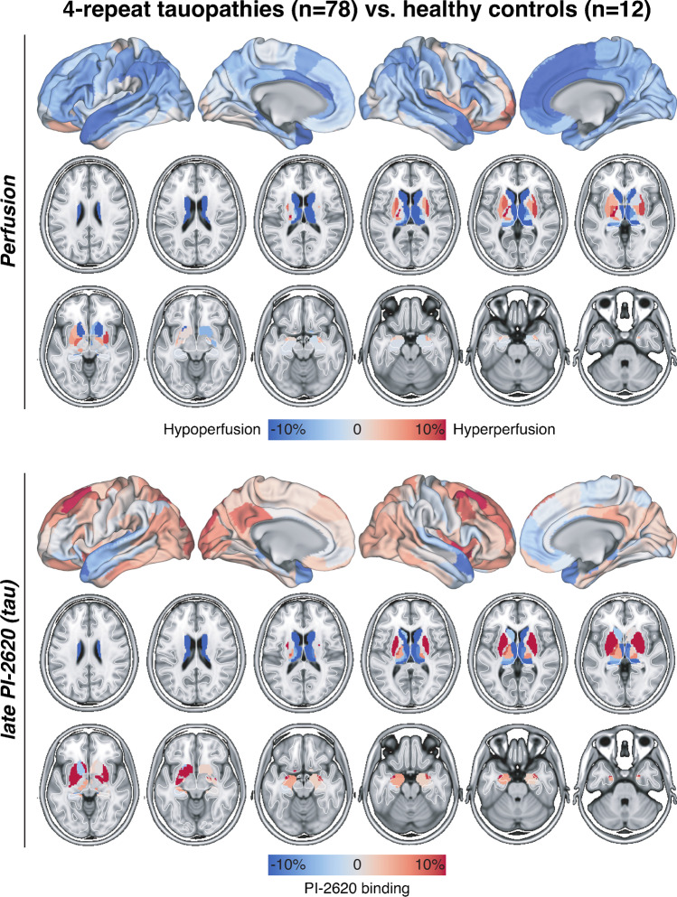
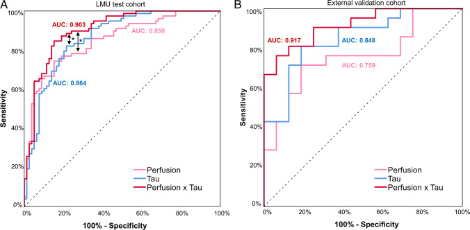
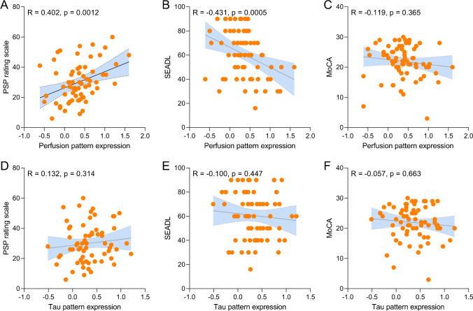
Results: Patients with 4RTs had significant hypoperfusion in 21/246 brain regions, most dominant in thalamus, caudate nucleus, and anterior cingulate cortex, fitting to the topology of the 4RT disease spectrum. However, single region hypoperfusion was not specific regarding the discrimination of patients with 4RTs against patients with other neurodegenerative diseases. In contrast, perfusion pattern expression showed promise for discrimination of patients with 4RTs from other neurodegenerative diseases (AUC: 0.850). Discrimination by the combined perfusion-tau pattern expression (AUC: 0.903) exceeded that of the sole tau pattern expression (AUC: 0.864) and the discriminatory power of the combined perfusion-tau pattern expression was replicated in the external dataset (AUC: 0.917). Perfusion but not tau pattern expression was associated with PSP rating scale (R = 0.402; p = 0.0012) and activities of daily living (R = - 0.431; p = 0.0005).
Conclusion: [18F]PI-2620 perfusion imaging mirrors known topology of regional hypoperfusion in 4RTs. Single region hypoperfusion is not specific for 4RTs, but perfusion pattern expression may provide an additive value for the discrimination of 4RTs from other neurodegenerative diseases and correlates closer with clinical severity than tau pattern expression.
Keywords: Neuronal injury; PET; Perfusion; Tau; [18F]PI-2620.
© 2022. The Author(s).
Conflict of interest statement
Johannes Levin reports speaker fees from Bayer Vital, Biogen, and Roche; consulting fees from Axon Neuroscience and Biogen; author fees from Thieme medical publishers and W. Kohlhammer GmbH medical publishers; non-financial support from Abbvie; and compensation for duty as part-time CMO from MODAG, all outside the submitted work. Andrew W. Stephens is a full-time employee of Life Molecular Imaging, GmbH. Thilo van Eimeren reports speaker/consultant fees from Eli Lilly, Shire, H. Lundbeck A/S, and Orion Corporation and author fees from Thieme medical publishers, all without conflict of interest with regard to the submitted work.
Figures

Comment on
-
Assessment of 18F-PI-2620 as a Biomarker in Progressive Supranuclear Palsy.JAMA Neurol. 2020 Nov 1;77(11):1408-1419. doi: 10.1001/jamaneurol.2020.2526. JAMA Neurol. 2020. PMID: 33165511 Free PMC article.
References
Publication types
MeSH terms
Substances
Grants and funding
LinkOut - more resources
Full Text Sources
Medical
Research Materials
Miscellaneous
