

Lung ultrasound in the follow-up of severe COVID-19 pneumonia: six months evaluation and comparison with CT
- PMID: 36103083
- PMCID: PMC9472735
- DOI: 10.1007/s11739-022-03084-9
Lung ultrasound in the follow-up of severe COVID-19 pneumonia: six months evaluation and comparison with CT
Abstract
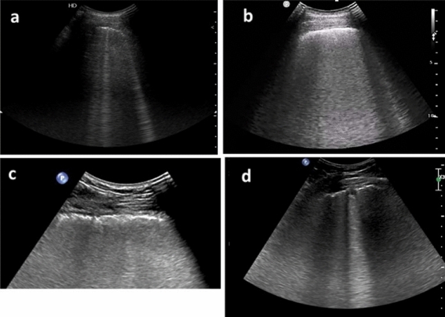
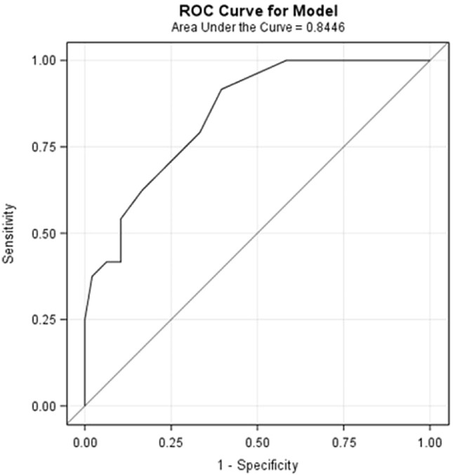
While lung ultrasonography (LUS) proved to be a useful diagnostic and prognostic tool in acute phase of COVID 19 pneumonia, its role in detecting long-term pulmonary sequelae has yet to be explored. In our prospective observational study we assessed the potential of LUS in detecting the presence of computed tomography (CT) fibrotic-like changes after 6 months from COVID-19 pneumonia. Patients who were discharged with a diagnosis of severe COVID-19 pneumonia were enrolled. After 6 months from hospital discharge they underwent LUS, chest CT scan and pulmonary function tests. A logistic regression analysis was performed to assess the association between presence of symptoms, LUS score and diffusing capacity for carbon monoxide (DLCO) at 6-month after hospital discharge and CT scan fibrotic-like changes. A second logistic model was performed to assess the value of some predefined baseline factors (age, sex, worst PaO2/FiO2, ventilator support, worst CRP value, worst D-dimer value and worst LUS score during hospitalization) to predict fibrotic-like changes on 6-month CT scan. Seventy-four patients were enrolled in the study. Twenty-four (32%) showed lung abnormalities suitable for fibrotic-like changes. At multivariate logistic regression analysis LUS score after 6 months from acute disease was significantly associated with fibrotic-like pattern on CT scan. The second logistic model showed that D-dimer value was the only baseline predictive variable of fibrotic-like changes at multivariate analysis. LUS performed after 6 months from severe COVID-19 pneumonia may be a promising tool for detection and follow-up of pulmonary fibrotic sequelae.
Keywords: COVID-19 pneumonia; Computed tomography; Lung ultrasound; Pulmonary fibrosis; Ultrasonography.
© 2022. The Author(s), under exclusive licence to Società Italiana di Medicina Interna (SIMI).
Conflict of interest statement
All authors declare that they have no conflicts of interest. Conflict of interest disclosure forms for each author are submitted separately as a single file.
Figures
Comment in
-
Can lung ultrasound replace CT scan in a 6-month follow-up of severe COVID-19 pneumonia? A brief commentary.Intern Emerg Med. 2023 Jun;18(4):977-978. doi: 10.1007/s11739-023-03210-1. Epub 2023 Apr 1. Intern Emerg Med. 2023. PMID: 37004666 Free PMC article. No abstract available.
References
-
- Ayoubkhani D, Gaughan C. Technical article : Updated estimates of the prevalence of post-acute symptoms among people with coronavirus (COVID-19) in the UK : 26 April 2020 to 1 August 2021. 2021; (April 2020):1–19. https://www.ons.gov.uk/peoplepopulationandcommunity/healthandsocialcare/....- Main%20points,data%20to%201%20August%202021
Publication types
MeSH terms
LinkOut - more resources
Full Text Sources
Medical
Research Materials
Miscellaneous
