

Rho Kinase Inhibitor Y27632 Improves Recovery After Spinal Cord Injury by Shifting Astrocyte Phenotype and Morphology via the ROCK/NF-κB/C3 Pathway
- PMID: 36103106
- PMCID: PMC9718714
- DOI: 10.1007/s11064-022-03756-0
Rho Kinase Inhibitor Y27632 Improves Recovery After Spinal Cord Injury by Shifting Astrocyte Phenotype and Morphology via the ROCK/NF-κB/C3 Pathway
Erratum in
-
Correction: Rho Kinase Inhibitor Y27632 Improves Recovery After Spinal Cord Injury by Shifting Astrocyte Phenotype and Morphology via the ROCK/NF-?B/C3 Pathway.Neurochem Res. 2022 Dec;47(12):3745-3746. doi: 10.1007/s11064-022-03764-0. Neurochem Res. 2022. PMID: 36244038 Free PMC article. No abstract available.
Abstract
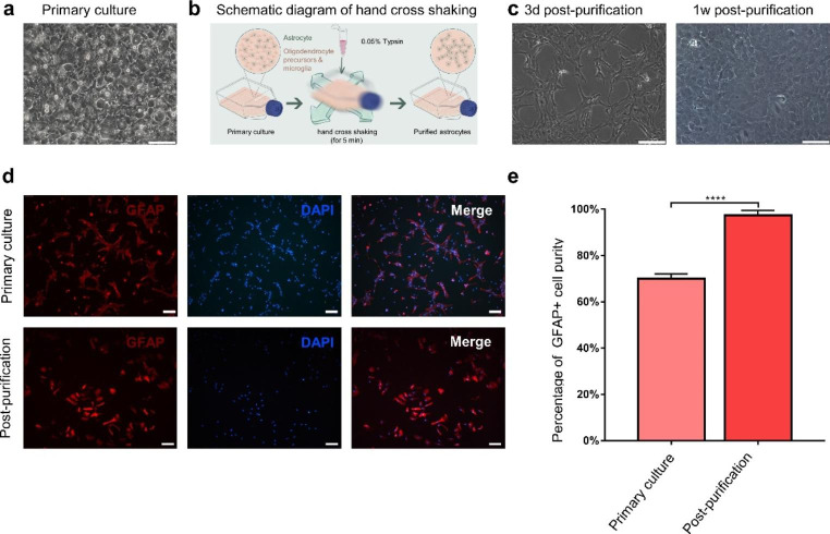
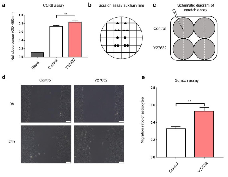
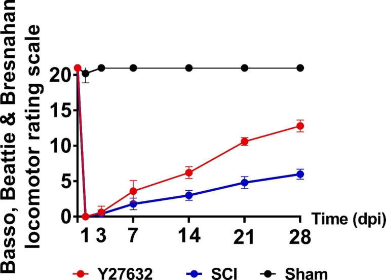
Spinal cord injury (SCI) usually results in loss or reduction in motor and sensory functions. Despite extensive research, no available therapy can restore the lost functions after SCI. Reactive astrocytes play a pivotal role in SCI. Rho kinase inhibitors have also been shown to promote functional recovery of SCI. However, the role of Rho kinase inhibitors in reactive astrocytic phenotype switch within SCI remains largely unexplored. In this study, astrocytes were treated with proinflammatory cytokines and/or the Rho kinase inhibitor Y27632. Concomitantly the phenotype and morphology of astrocytes were examined. Meanwhile, the SCI model of SD rats was established, and nerve functions were evaluated following treatment with Y27632. Subsequently, the number of A1 astrocytes in the injured area was observed and analyzed. Eventually, the expression levels of nuclear factor kappa B (NF-κB), C3, and S100A10 were measured. The present study showed that the Rho kinase inhibitor Y27632 improved functional recovery of SCI and elevated the proliferation and migration abilities of the astrocytes. In addition, Y27632 treatment initiated the switch of astrocytes morphology from a flattened shape to a process-bearing shape and transformed the reactive astrocytes A1 phenotype to an A2 phenotype. More importantly, further investigation suggested that Y27632 was actively involved in promoting the functional recovery of SCI in rats by inhabiting the ROCK/NF-κB/C3 signaling pathway. Together, Rho kinase inhibitor Y27632 effectively promotes the functional recovery of SCI by shifting astrocyte phenotype and morphology. Furthermore, the pro-regeneration event is strongly associated with the ROCK/NF-κB/C3 signal pathway.
Keywords: Astrocyte; C3; NF-κB; Rho kinase inhibitor; S100A10; Spinal cord injury.
© 2022. The Author(s).
Conflict of interest statement
The authors have no relevant financial or non-financial interests to disclose.
Figures


Similar articles
-
Synergistic effects of tetramethylpyrazine and astragaloside IV on spinal cord injury via alteration of astrocyte A1/A2 polarization through the Sirt1-NF-κB pathway.Int Immunopharmacol. 2024 Apr 20;131:111686. doi: 10.1016/j.intimp.2024.111686. Epub 2024 Mar 10. Int Immunopharmacol. 2024. PMID: 38461631
-
Geniposide exerts protective effects on spinal cord injury in rats by inhibiting the IKKs/NF-κB signaling pathway.Int Immunopharmacol. 2021 Nov;100:108158. doi: 10.1016/j.intimp.2021.108158. Epub 2021 Sep 20. Int Immunopharmacol. 2021. PMID: 34555642
-
Combined application of Rho-ROCKII and GSK-3β inhibitors exerts an improved protective effect on axonal regeneration in rats with spinal cord injury.Mol Med Rep. 2016 Dec;14(6):5180-5188. doi: 10.3892/mmr.2016.5918. Epub 2016 Nov 1. Mol Med Rep. 2016. PMID: 27840930 Free PMC article.
-
Rho-ROCK inhibition in the treatment of spinal cord injury.World Neurosurg. 2014 Sep-Oct;82(3-4):e535-9. doi: 10.1016/j.wneu.2013.01.009. Epub 2013 Jan 5. World Neurosurg. 2014. PMID: 23298675 Review.
-
Exploring the potential of RhoA inhibitors to improve exercise-recoverable spinal cord injury: A systematic review and meta-analysis.J Chem Neuroanat. 2021 Jan;111:101879. doi: 10.1016/j.jchemneu.2020.101879. Epub 2020 Nov 13. J Chem Neuroanat. 2021. PMID: 33197553
Cited by
-
The RhoA-ROCK1/ROCK2 Pathway Exacerbates Inflammatory Signaling in Immortalized and Primary Microglia.Cells. 2023 May 11;12(10):1367. doi: 10.3390/cells12101367. Cells. 2023. PMID: 37408199 Free PMC article.
-
Traumatic Spinal Cord Injury: Review of the Literature.J Clin Med. 2025 May 22;14(11):3649. doi: 10.3390/jcm14113649. J Clin Med. 2025. PMID: 40507410 Free PMC article. Review.
-
The potential role of RhoA/ROCK-inhibition on locomotor recovery after spinal cord injury: a systematic review of in-vivo studies.Spinal Cord. 2025 Mar;63(3):95-126. doi: 10.1038/s41393-025-01064-2. Epub 2025 Feb 16. Spinal Cord. 2025. PMID: 39956860
-
Ruxolitinib improves the inflammatory microenvironment, restores glutamate homeostasis, and promotes functional recovery after spinal cord injury.Neural Regen Res. 2024 Nov 1;19(11):2499-2512. doi: 10.4103/NRR.NRR-D-23-01863. Epub 2024 Jan 31. Neural Regen Res. 2024. PMID: 38526286 Free PMC article.
-
Development of Tissue-Engineered Model of Fibrotic Scarring after Spinal Cord Injury to Study Astrocyte Activation and Neurite Outgrowth In Vitro.ACS Biomater Sci Eng. 2024 Oct 14;10(10):6545-6557. doi: 10.1021/acsbiomaterials.4c01100. Epub 2024 Sep 11. ACS Biomater Sci Eng. 2024. PMID: 39259933 Free PMC article.
References
-
- Virchow R. Gesammelte Abhandlungen Zur Wissenschaftlichen Medizin. Frankf Am Taf. 1856;20:1–1024.
MeSH terms
Substances
Grants and funding
LinkOut - more resources
Full Text Sources
Medical
Miscellaneous
