. 2022 Sep 14;17(9):e0261805.
doi: 10.1371/journal.pone.0261805. eCollection 2022.

Gross anatomy of the gluteal and posterior thigh muscles in koalas based on their innervations

Affiliations

Gross anatomy of the gluteal and posterior thigh muscles in koalas based on their innervations

Sayaka Tojima et al. PLoS One. .

Abstract

Morphological and functional comparison of convergently-evolved traits in marsupials and eutherians is an important aspect of studying adaptive divergence in mammals. However, the anatomy of marsupials has been particularly difficult to evaluate for multiple reasons. First, previous studies on marsupial anatomy are often uniformly old and non-exhaustive. Second, muscle identification was historically based on muscle attachment sites, but attachment sites have since been declared insufficient for muscle identification due to extensive interspecific variation. For example, different names have been used for muscles that are now thought to be equivalent among several different species, which causes confusion. Therefore, descriptions of marsupial muscles have been inconsistent among previous studies, and their anatomical knowledge itself needs updating. In this study, the koala was selected as the representative marsupial, in part because koala locomotion may comprise primate (eutherian)-like and marsupial-like mechanics, making it an interesting phylogenetic group for studying adaptive divergence in mammals. Gross dissection of the lower limb muscles (the gluteal and the posterior thigh regions) was performed to permit precise muscle identification. We first resolved discrepancies among previous studies by identifying muscles according to their innervation; this recent, more reliable technique is based on the ontogenetic origin of the muscle, and it allows for comparison with other taxa (i.e., eutherians). We compared our findings with those of other marsupials and arboreal primates and identified traits common to both arboreal primates and marsupials as well as muscle morphological features unique to koalas.


Conflict of interest statement

The authors have declared that no competing interests exist.

Figures

Fig 1
Fig 1. Superficial layer muscles of gluteal and posterior femoral regions in a koala right hind limb.
(a) The superficial gluteal muscles, the gluteus superficialis (GSu) and superficial part of the gluteus medius (GMS), and some cutaneous nerves are shown. (b) The thigh flexors and some cutaneous nerves are shown. (c) Illustrated schematic drawing of the superficial layer muscles of gluteal and posterior femoral regions. Abbreviations: BF, biceps femoris; GF, gluteal fascia; GMS, superficial layer of the gluteus medius; GR, gracilis; GrT, greater trochanter; GSu, gluteus superficialis; HB, hamstring bundle; ICN, inferior cluneal nerve; MCN, middle cluneal nerve; PFCN, posterior femoral cutaneous nerve; SCN, superior cluneal nerve; ST, semitendinosus; STR, semitendinosus raphe; VL, vastus lateralis.
Fig 2
Fig 2. Deep layer muscles of gluteal and posterior femoral regions in a koala right hind-limb.
(a) The deep gluteus muscles and several nerves including muscular branches are shown. (a) The deep tibial nerve (DTN) and deep gluteus muscles deep to the piriformis (PI) and ischiofemoralis (IF) are shown. (b) The innervating branches to the thigh flexors are shown. (c) Schematic drawing of deep gluteal muscles, thigh flexors, and innervating branches. White arrowheads indicate the innervating branch from the deep tibial nerve (DTN) to the hamstring bundle (HB). Abbreviations: AM, adductor magnus; BF, biceps femoris; DTN, deep tibial nerve; GlT, gluteal tuberosity; GMD, deep layer of the gluteus medius; GMS, superficial layer of the gluteus medius; GrT, greater trochanter; GSu, gluteus superficialis; HB, hamstring bundle; IF, ischiofemoralis; IG, inferior gemellus; IGN, inferior gluteal nerve; PFCN, posterior femoral cutaneous nerve; PI, piriformis; SG, superior gemellus; SGN, superior gluteal nerve; SM, semimembranosus; SN, sciatic nerve; ST, semitendinosus; VL, vastus lateralis.
Fig 3
Fig 3. Deepest layer muscles of gluteus region and its innervations in koala right hip.
(a) The superficial and deep layers of the gluteus medius (GMS and GMD), gluteus minimus (GMi), and the innervating branch from the superior gluteal nerve (SGN) are shown. (b) Gemellus and internal obturator muscles and supplying branch from the deep tibial nerve (DTN) are shown. (c) Schematic drawing of the deepest layer of gluteus muscles and its innervations. Abbreviations: DTN, deep tibial nerve; GlT, gluteal tuberosity; GMD, deep layer of the gluteus medius; GMi, gluteus minimus; GMS, superficial layer of the gluteus medius; GrT, greater trochanter; IG, inferior gemellus; IGN, inferior gluteal nerve; IO, internal obturator; PI, piriformis; SG, superior gemellus; SGN, superior gluteal nerve; SN, sciatic nerve.
Fig 4
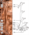
Fig 4. Right lumbosacral plexus in koalas.
Photograph and illustrated corresponding schematic drawing. White and black asterisks indicate branches forming the posterior femoral cutaneous nerve and the innervating branch to the piriformis and ischiofemoralis after leaving the greater sciatic foramen. Abbreviations: DTN, deep tibial nerve; FN, femoral nerve; GSF, greater sciatic foramen; IGN, inferior gluteal nerve; LT, lumbosacral trunk; ON, obturator nerve; PN, pudendal nerve; SGN, superior gluteal nerve; SN, sciatic nerve.

References

    1. Nilsson MA, Churakov G, Sommer M, Tran NV, Zemann A, et al.. Tracking marsupial evolution using archaic genomic retroposon insertions. PLoS Biol 2010; 8(7): e1000436. doi: 10.1371/journal.pbio.1000436 - DOI - PMC - PubMed
    1. Schmitt D., Lemelin P. Origins of primate locomotion: gait mechanics of the woolly opossum. Am J Phys Anthropol 2002; 118, 231–238. doi: 10.1002/ajpa.10048 - DOI - PubMed
    1. Lemelin P., Schmitt D., Cartmill M. Footfall patterns and interlimb co-ordination in opossums (Family Didelphidae): evidence for the evolution of diagonal-sequence walking gaits in primates. J Zool Lond 2003; 260, 423–429.
    1. Gaschk J. L., Frère C. H., Clemente C. J. Quantifying koala locomotion strategies: implications for the evolution of arborealism in marsupials. J Exp Biol 2019; 222, jeb207506. doi: 10.1242/jeb.207506 - DOI - PubMed
    1. Smith-Paredes D, Vegara-Cereghino M, Lord A, Moses MM, Behringer RR, Bhullar BA. Embryonic muscle splitting patterns reveal homologies of amniote forelimb muscles. Nature Eco Evo 2022; 6: 604–613. doi: 10.1038/s41559-022-01699-x - DOI - PMC - PubMed

Publication types