. 2022 Sep 20;119(38):e2208337119.
doi: 10.1073/pnas.2208337119. Epub 2022 Sep 14.

Synaptotagmin rings as high-sensitivity regulators of synaptic vesicle docking and fusion

Affiliations

Synaptotagmin rings as high-sensitivity regulators of synaptic vesicle docking and fusion

Jie Zhu et al. Proc Natl Acad Sci U S A. .

Abstract

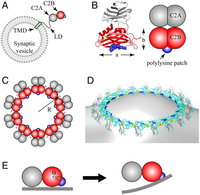
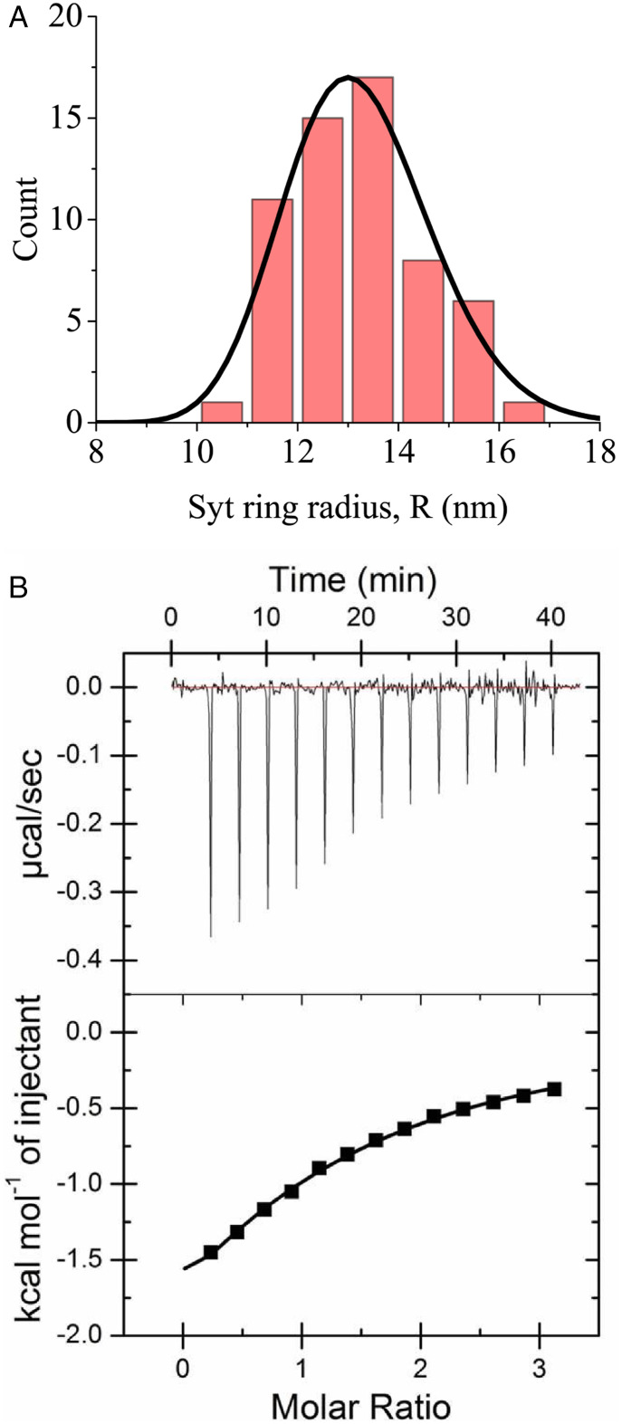
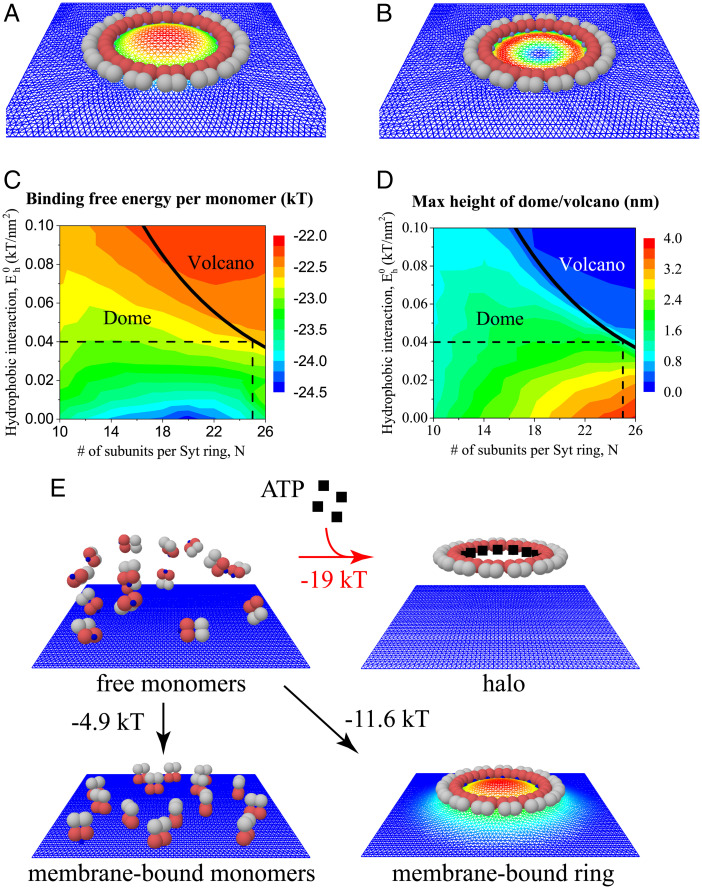
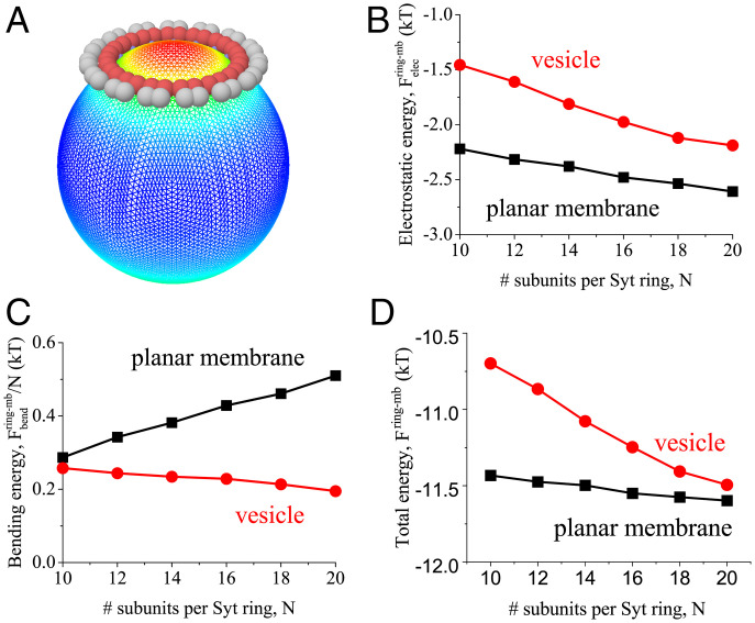
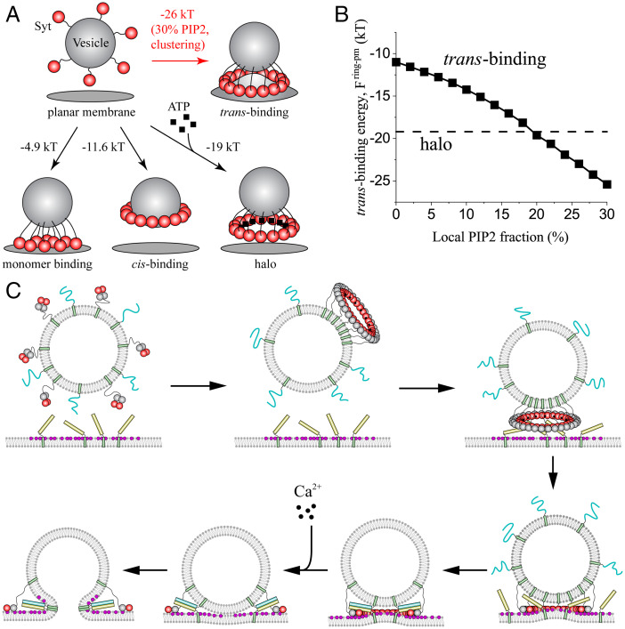
Synchronous release at neuronal synapses is accomplished by a machinery that senses calcium influx and fuses the synaptic vesicle and plasma membranes to release neurotransmitters. Previous studies suggested the calcium sensor synaptotagmin (Syt) is a facilitator of vesicle docking and both a facilitator and inhibitor of fusion. On phospholipid monolayers, the Syt C2AB domain spontaneously oligomerized into rings that are disassembled by Ca2+, suggesting Syt rings may clamp fusion as membrane-separating "washers" until Ca2+-mediated disassembly triggers fusion and release [J. Wang et al., Proc. Natl. Acad. Sci. U.S.A. 111, 13966-13971 (2014)].). Here, we combined mathematical modeling with experiment to measure the mechanical properties of Syt rings and to test this mechanism. Consistent with experimental results, the model quantitatively recapitulates observed Syt ring-induced dome and volcano shapes on phospholipid monolayers and predicts rings are stabilized by anionic phospholipid bilayers or bulk solution with ATP. The selected ring conformation is highly sensitive to membrane composition and bulk ATP levels, a property that may regulate vesicle docking and fusion in ATP-rich synaptic terminals. We find the Syt molecules hosted by a synaptic vesicle oligomerize into a halo, unbound from the vesicle, but in proximity to sufficiently phosphatidylinositol 4,5-bisphosphate (PIP2)-rich plasma membrane (PM) domains, the PM-bound trans Syt ring conformation is preferred. Thus, the Syt halo serves as landing gear for spatially directed docking at PIP2-rich sites that define the active zones of exocytotic release, positioning the Syt ring to clamp fusion and await calcium. Our results suggest the Syt ring is both a Ca2+-sensitive fusion clamp and a high-fidelity sensor for directed docking.

Keywords: membrane fusion; neurotransmitter release; synapse; synaptotagmin; vesicle docking.


Conflict of interest statement

The authors declare no competing interest.

Figures

Fig. 1.
Fig. 1.
Coarse-grained model of Syt C2AB domain and oligomeric Syt rings. (A) Synaptic vesicles carry ∼15 copies of Syt, with each comprising a TMD, a flexible juxtamembrane LD, and the C2A and C2B globular domains (schematic, not to scale). The polylysine patch (blue) on C2B mediates Ca2+-independent binding to anionic membranes. (B) Crystal structure of Syt (PDB ID: 2R83) (Left) and our coarse-grained representation (Right). Each C2 domain is represented as two overlapping beads, radius Rbead, giving length a=5 nm and width b=3 nm. The positively charged polylysine patch (blue) on C2B is treated as a point charge. (C) Coarse-grained model representation of a Syt ring of radius R, Top view. (D) Rendition of a Syt ring bound to a membrane, based on EM reconstruction of Syt oligomers assembled on phospholipid monolayer tubes (43) (cyan, C2B; gray, C2A; blue, polylysine patch; yellow, Ca2+ binding loops on C2B). (E) C2AB subunit in a ring interacting with planar membrane (Side view). The polylysine patch lies below the plane of the ring, at an angle θ=30° suggested by EM reconstruction, D. In the ring, the C2AB unit cannot rotate downward, so electrostatic attraction bends the charged membrane up toward the patch (Right).
Fig. 2.
Fig. 2.
(A) Experimental Syt ring size distribution in bulk solution with 1 mM Mg-ATP and 15 mM KCl, from electron micrographs (radius R is defined in Fig. 1C). Mean radius is 13.1 nm ± 1.4 nm (SD). Solid line represents best fit theoretical distribution (Eq. 1), yielding spontaneous radius of curvature R0=13 nm and persistence length lp=170 nm (Table 1). (B) Syt-ATP binding measured by ITC. The measured parameters are as follows: ΔH = −4.4 ± 0.7 kcal/mol, dissociation constant kdsyt-ATP=105 ±13 µM.
Fig. 3.
Fig. 3.
Model results for binding of Syt rings on carbon-supported monolayers and on planar bilayer membranes. (A and B) Simulation snapshots of equilibrium configurations of a Syt ring of N=16 subunits binding a monolayer with 40% PS, 60% PC in 15 mM salt solution (conditions as in ref. 43). Warmer colors denote greater monolayer elevations. (A) Monolayer deforms into a dome shape for lower monolayer-carbon hydrophobic interaction energy, Eh0=0.05 kT/nm2. (B) Volcano shape results for larger interaction, Eh0=0.15 kT/nm2. (C and D) Monolayer shape (dome or volcano) and binding free energy per monomer (C) or maximum monolayer height (D) versus N, the number of Syt subunits per ring, and Eh0, the monolayer-carbon binding energy density. In the experiments of ref. , the dome–volcano transition occurred at N25, identifying Eh0=0.04 kT/nm2 (dashed lines). (E) Possible states for Syt monomers in solution near a planar bilayer membrane with overall composition typical of a plasma membrane, with composition assumed homogenous (Table 2). Energies of formation are per Syt monomer, relative to a reference state with 15 free Syt monomers in solution at physiological salt conditions, 140 mM (Top Left). With ATP, the preferred state is the Syt ring in solution (halo), stabilized by the substantial Syt-ATP binding energy, ϵsytATP9.7 kT Without ATP, the Syt ring oligomerizes on the membrane, deforming it into a dome (simulation snapshot, warmer colors denote greater elevation).
Fig. 4.
Fig. 4.
Membrane curvature and charge density promote binding of Syt rings. Model results for binding of Syt rings at physiological salt, 140 mM, to a 20-nm-radius lipid vesicle with composition representative of synaptic vesicles (15% PS, Table 2). Vesicle binding energies are compared with binding energies to planar membranes with lipid composition representative of a plasma membrane (Table 2). Energies are per Syt subunit. (A) Simulation snapshot of a Syt ring of N=15 subunits binding and dimpling the vesicle, with a mild version of the dome seen on planar bilayers. Warmer colors denote greater distance from vesicle center. (BD) Model-predicted free energies when a Syt ring binds a vesicle or a planar membrane versus number of Syt monomers per ring. (B) The attractive electrostatic energy is greater for the plasma membrane due to its higher anionic lipid charge density. (C) Vesicle binding incurs less bending energy, due to vesicle curvature. (D) The net binding energy favors the plasma membrane since the electrostatic advantage exceeds the bending disadvantage.
Fig. 5.
Fig. 5.
(A) When a synaptic vesicle bearing 15 Syt molecules approaches the plasma membrane, 4 final states are possible. The model-predicted free energies per molecule are shown. Vesicle size, lipid composition, and salt conditions are as for Fig. 4. At PIP2 clustering sites with sufficiently high local PIP2 concentration, trans-binding of a Syt ring is preferred (e.g., 30% PIP2, as shown) to the ATP-stabilized halo, the cis-bound ring, or unoligomerized membrane-bound monomers. (B) Model calculations of trans-binding energy of a Syt ring to the plasma membrane versus local PIP2 concentration. For [PIP2] >18%, trans-binding is favored over the halo, while halos are preferred for [PIP2] < 18%. (C) Model of Syt-mediated regulation of neurotransmitter release, with the Syt ring serving both as a discriminating sensor and as a spacer clamping fusion. In the ATP-rich synaptic terminal, the 15 SytC2AB monomers carried by a synaptic vesicle form a halo (unbound ring) in preference to a ring bound to the PIP2-free vesicle (cis-binding). The oligomeric nature of the Syt ring endows it with high sensitivity to PIP2 levels in the plasma membrane, and trans-binding to the PM is preferred to the halo only for PIP2 content above a threshold. Thus, the Syt halo is spatially directed to dock the vesicle at PIP2-rich sites that colocalize with the t-SNARE Syntaxin (yellow). Docking positions the Syt ring to clamp fusion by spacing the vesicle and PMs and by restraining the SNARE proteins to lie partially within the Syt ring, consistent with measured crystal structures of the Syt-SNARE complex (23, 24). Ca2+ influx disassembles the ring, releasing SNAREs to fuse the membranes rapidly and release neurotransmitters through the fusion pore.

References

    1. Takamori S., et al. , Molecular anatomy of a trafficking organelle. Cell 127, 831–846 (2006). - PubMed
    1. Mutch S. A., et al. , Protein quantification at the single vesicle level reveals that a subset of synaptic vesicle proteins are trafficked with high precision. J. Neurosci. 31, 1461–1470 (2011). - PMC - PubMed
    1. Wilhelm B. G., et al. , Composition of isolated synaptic boutons reveals the amounts of vesicle trafficking proteins. Science 344, 1023–1028 (2014). - PubMed
    1. Chapman E. R., How does synaptotagmin trigger neurotransmitter release? Annu. Rev. Biochem. 77, 615–641 (2008). - PubMed
    1. Chapman E. R., Synaptotagmin: A Ca(2+) sensor that triggers exocytosis? Nat. Rev. Mol. Cell Biol. 3, 498–508 (2002). - PubMed

Publication types

MeSH terms

LinkOut - more resources