Review
. 2022 Sep 13;55(9):1564-1580.
doi: 10.1016/j.immuni.2022.08.010.

Biology of lung macrophages in health and disease

Affiliations
Review

Biology of lung macrophages in health and disease

Helena Aegerter et al. Immunity. .

Abstract

Tissue-resident alveolar and interstitial macrophages and recruited macrophages are critical players in innate immunity and maintenance of lung homeostasis. Until recently, assessing the differential functional contributions of tissue-resident versus recruited macrophages has been challenging because they share overlapping cell surface markers, making it difficult to separate them using conventional methods. This review describes how scRNA-seq and spatial transcriptomics can separate these subpopulations and help unravel the complexity of macrophage biology in homeostasis and disease. First, we provide a guide to identifying and distinguishing lung macrophages from other mononuclear phagocytes in humans and mice. Second, we outline emerging concepts related to the development and function of the various lung macrophages in the alveolar, perivascular, and interstitial niches. Finally, we describe how different tissue states profoundly alter their functions, including acute and chronic lung disease, cancer, and aging.

Keywords: alveolar macrophages; interstitial macrophages; lung disease; pulmonary; recruited macrophages.


Conflict of interest statement

Declaration of interests The authors declare no competing interests.

Figures
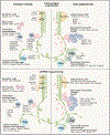
Figure 1.
Figure 1.. Cell surface protein and scRNA-seq identification of mononuclear phagocytes in the mouse lung
Top panel: CD45+ mononuclear phagocytes represented in the figure have varying surface expression of CD11c and CD11b. Prior to defining mononuclear phagocyte cell types using protein-based approaches such as flow cytometry, to avoid contamination, other immune cell types need to be excluded using lineage markers expressed on lymphoid cells (NK1.1, CD19, and CD3), neutrophils (Ly6G), and eosinophils (SiglecF+, SSChi, CD11cCD64). CD88 versus CD26 distinguishes monocytes and macrophages from cDCs. Note that monocytes do express a little CD26, but cDCs are CD88 negative. Activated monocytes and macrophages demonstrate high CD88 expression. AMs and IMs highly express MerTK (R&D: Polyclonal Goat IgG #AF591) and CD64 compared with recently recruited monocytes, which have low expression of MerTK, CD64, and F4/80 and varying surface expression of MHCII, CD11c, and Ly6C. Ly6C expression on recruited monocytes is lost over time. CD206 expression on in vivo macrophage does not define M2 macrophages since all AMs highly express CD206 and are not all M2-like. IMs express CD206 to a high and intermediate degree, as do cDC2s, recruited monocytes and macrophages. The two extreme profiles of IMs are represented here, although IMs, currently, based on the markers represented, are more a gradient of cell types than two clearly defined cell types. Similarly, cDC2 has multiple subtypes such as CD301+ and CD301 cDC2 (not represented here). Bottom panel: some commonly used genes to identify murine mononuclear phagocytes in scRNA-seq datasets. Like protein cell surface expression, CD88 versus CD26 gene expression help distinguish monocytes and macrophages from DCs. cDC, conventional dendritic cells; TRAM, tissue resident alveolar macrophage; IM, interstitial macrophage; moDC, monocyte-derived DC; recAM, recruited alveolar macrophages; recMac, recruited macrophages; Inf-cDC, inflammatory conventional DCs.
Figure 2.
Figure 2.. Development of alveolar macrophages from fetal precursors
Around embryonic (E) day 18.5, fetal monocytes exit the lung vasculature and under the influence of autocrine TGF-β turn on a macrophage differentiation program characterized by expression of macrophage lineage-determining transcription factors. These cells next migrate to the alveolar niche where around the day of birth (DOB), they develop into immature TRAM, under the influence of granulocyte-macrophage colony-stimulating factor (GM-CSF) produced by type II alveolar epithelial cells and helped by macrophage-intrinsic transforming growth factor beta (TGF-β). This induces the expression of the transcription factor PPARγ that controls a large part of the AM-specific transcriptome. Mature TRAMs also contain transcription factors that allow for AM self-renewal. AM identity is instructed by other epithelial-cell-derived molecules like surfactant proteins A and D (SP-A and SP-D), TGF-β, and isthmin.
Figure 3.
Figure 3.. Metabolism of alveolar macrophages in homeostasis and infection
Steady-state AMs exist in an environment of low glucose and other environmental signals that instruct their metabolism and inflammatory output. PPARγ is a key AM transcription factor induced by GM-CSF, which regulates lipid metabolism, a key feature of AM functionality. In contrast, recAMs develop in an environment of higher glucose accessibility, and due to an altered origin or altered environmental signals, remain in an altered metabolic state characterized by high HIF1a that results in increased of inflammatory cytokines and nitric oxide.
Figure 4.
Figure 4.. Involvement of lung macrophages in development of lung fibrosis
In homeostasis, TRAMs clear out apoptotic cells and other debris and, in this process, produce pro-repair genes like amphiregulin and TGF-β that is necessary for normal tissue homeostasis and repair. In fibrosis developing in response to injury, or due to an idiopathic epithelial injury, however monocytes are recruited and as these develop into recMacs or recAMs, they become programmed to contribute to fibrosis by producing profibrotic growth factors and chemokines, matrix metalloproteinases, matrix components, and other mediators. These cause activation of type 2 epithelial cells, which become hyperplastic, and activation of myofibroblasts that produce too much collagens. However, both epithelial cells and myofibroblasts then produce cytokines that can activate the recMacs to contribute further to fibrosis and can activate other niche cells to produce more collagens. This becomes a self-sustaining functional unit in progressive fibrosis.

References

    1. Aegerter H, Kulikauskaite J, Crotta S, Patel H, Kelly G, Hessel EM, Mack M, Beinke S, and Wack A (2020). Influenza-induced monocyte-derived alveolar macrophages confer prolonged antibacterial protection. Nat. Immunol 21, 145–157. - PMC - PubMed
    1. Angelidis I, Simon LM, Fernandez IE, Strunz M, Mayr CH, Greiffo FR, Tsitsiridis G, Ansari M, Graf E, Strom TM, et al. (2019). An atlas of the aging lung mapped by single cell transcriptomics and deep tissue proteomics. Nat. Commun 10, 963. - PMC - PubMed
    1. Antonelli LRV, Gigliotti Rothfuchs A, Gonçalves R, Roffê E, Cheever AW, Bafica A, Salazar AM, Feng CG, and Sher A (2010). Intranasal Poly-IC treatment exacerbates tuberculosis in mice through the pulmonary recruitment of a pathogen-permissive monocyte/macrophage population. J. Clin. Invest 120, 1674–1682. - PMC - PubMed
    1. Arafa EI, Shenoy AT, Barker KA, Etesami NS, Martin IM, Lyon De Ana C, Na E, Odom CV, Goltry WN, Korkmaz FT, et al. (2022). Recruitment and training of alveolar macrophages after pneumococcal pneumonia. JCI Insight 7, e150239. - PMC - PubMed
    1. Aran D, Looney AP, Liu L, Wu E, Fong V, Hsu A, Chak S, Naikawadi RP, Wolters PJ, Abate AR, et al. (2019). Reference-based analysis of lung single-cell sequencing reveals a transitional profibrotic macrophage. Nat. Immunol 20, 163–172. - PMC - PubMed

Publication types