

Generation and in vivo validation of an IL-12 fusion protein based on a novel anti-human FAP monoclonal antibody
- PMID: 36104101
- PMCID: PMC9476130
- DOI: 10.1136/jitc-2022-005282
Generation and in vivo validation of an IL-12 fusion protein based on a novel anti-human FAP monoclonal antibody
Abstract
Background: In this study, we describe the generation of a fully human monoclonal antibody (named '7NP2') targeting human fibroblast activation protein (FAP), an antigen expressed in the microenvironment of different types of solid neoplasms.
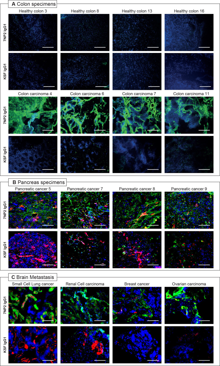
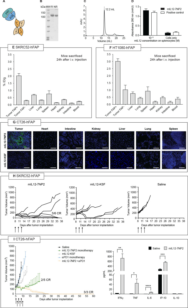
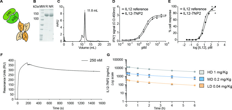
Methods: 7NP2 was isolated from a synthetic antibody phage display library and was improved by one round of mutagenesis-based affinity maturation. The tumor recognition properties of the antibody were validated by immunofluorescence procedures performed on cancer biopsies from human patients. A fusion protein consisting of the 7NP2 antibody linked to interleukin (IL)-12 was generated and the anticancer activity of the murine surrogate product (named mIL12-7NP2) was evaluated in mouse models. Furthermore, the safety of the fully human product (named IL12-7NP2) was evaluated in Cynomolgus monkeys.
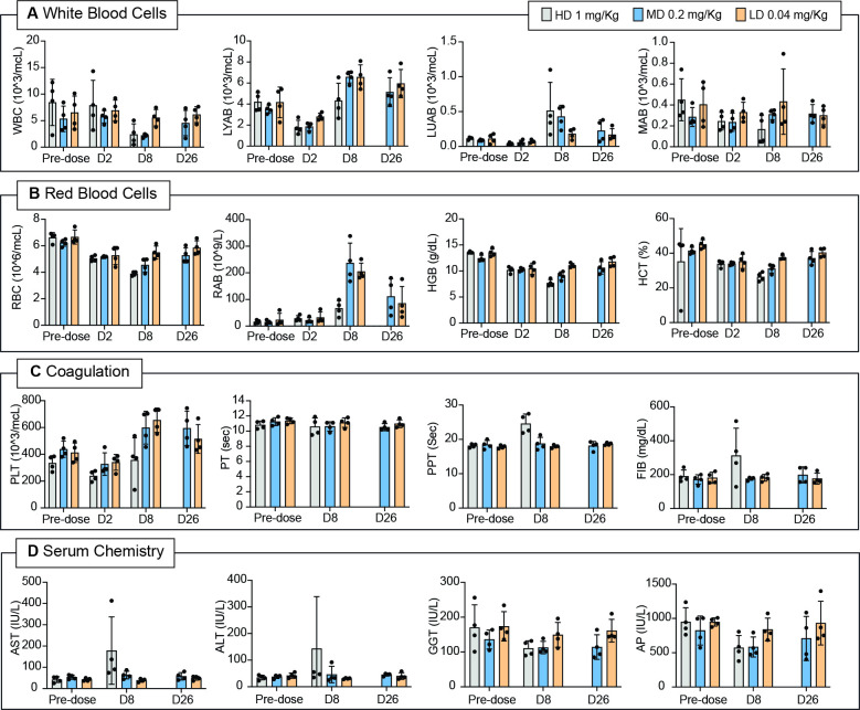
Results: Biodistribution analysis in tumor-bearing mice confirmed the ability of the product to selectively localize to solid tumors while sparing healthy organs. Encouraged by these results, therapy studies were conducted in vivo, showing a potent antitumor activity in immunocompetent and immunodeficient mouse models of cancer, both as single agent and in combination with immune checkpoint inhibitors. The fully human product was tolerated when administered to non-human primates.
Conclusions: The results obtained in this work provided a rationale for future clinical translation activities using IL12-7NP2.
Keywords: antibodies, neoplasm; antigens; cytokines; immunotherapy.
© Author(s) (or their employer(s)) 2022. Re-use permitted under CC BY-NC. No commercial re-use. See rights and permissions. Published by BMJ.
Conflict of interest statement
Competing interests: DN is a co-founder and shareholder of Philogen (www.philogen.com), a Swiss- Italian Biotech company that operates in the field of ligand-based pharmacodelivery. LN, FP, AE, NF, EPuc, CDN, EPro, RC, MM, and RD are employees of Philochem AG, daughter company of Philogen acting as discovery unit of the group.
Figures
Similar articles
-
Novel human monoclonal antibodies specific to the alternatively spliced domain D of Tenascin C efficiently target tumors in vivo.MAbs. 2020 Jan-Dec;12(1):1836713. doi: 10.1080/19420862.2020.1836713. MAbs. 2020. PMID: 33136526 Free PMC article.
-
The antibody-based delivery of interleukin-12 to the tumor neovasculature eradicates murine models of cancer in combination with paclitaxel.Clin Cancer Res. 2012 Aug 1;18(15):4092-103. doi: 10.1158/1078-0432.CCR-12-0282. Epub 2012 Jun 12. Clin Cancer Res. 2012. PMID: 22693354
-
Generation and in vivo characterization of a novel high-affinity human antibody targeting carcinoembryonic antigen.MAbs. 2023 Jan-Dec;15(1):2217964. doi: 10.1080/19420862.2023.2217964. MAbs. 2023. PMID: 37243574 Free PMC article.
-
Cytokine, chemokine, and co-stimulatory fusion proteins for the immunotherapy of solid tumors.Handb Exp Pharmacol. 2008;(181):291-328. doi: 10.1007/978-3-540-73259-4_13. Handb Exp Pharmacol. 2008. PMID: 18071951 Review.
-
Antibody-Cytokine Fusions: Versatile Products for the Modulation of Anticancer Immunity.Cancer Immunol Res. 2019 Mar;7(3):348-354. doi: 10.1158/2326-6066.CIR-18-0622. Cancer Immunol Res. 2019. PMID: 30824549 Free PMC article. Review.
Cited by
-
From basic research to clinical application: targeting fibroblast activation protein for cancer diagnosis and treatment.Cell Oncol (Dordr). 2024 Apr;47(2):361-381. doi: 10.1007/s13402-023-00872-z. Epub 2023 Sep 19. Cell Oncol (Dordr). 2024. PMID: 37726505 Review.
-
An Engineered IFNγ-Antibody Fusion Protein with Improved Tumor-Homing Properties.Pharmaceutics. 2023 Jan 22;15(2):377. doi: 10.3390/pharmaceutics15020377. Pharmaceutics. 2023. PMID: 36839699 Free PMC article.
-
4-1BB agonist targeted to fibroblast activation protein α synergizes with radiotherapy to treat murine breast tumor models.J Immunother Cancer. 2025 Feb 11;13(2):e009852. doi: 10.1136/jitc-2024-009852. J Immunother Cancer. 2025. PMID: 39933836 Free PMC article.
-
An Antibody Targeting Fibroblast Activation Protein Simultaneously Fused to Interleukin-2 and Tumor Necrosis Factor Selectively Localizes to Neoplastic Lesions.Antibodies (Basel). 2023 Apr 14;12(2):29. doi: 10.3390/antib12020029. Antibodies (Basel). 2023. PMID: 37092450 Free PMC article.
-
Targeting PD-1+ T cells with small-format immunocytokines enhances IL-12 antitumor activity.Mol Ther. 2025 Jan 8;33(1):297-316. doi: 10.1016/j.ymthe.2024.11.027. Epub 2024 Nov 19. Mol Ther. 2025. PMID: 39563030
References
MeSH terms
Substances
LinkOut - more resources
Full Text Sources
Medical
Miscellaneous