. 2022 Sep;10(9):e005116.
doi: 10.1136/jitc-2022-005116.

FBW7-mediated ubiquitination and destruction of PD-1 protein primes sensitivity to anti-PD-1 immunotherapy in non-small cell lung cancer

Affiliations

FBW7-mediated ubiquitination and destruction of PD-1 protein primes sensitivity to anti-PD-1 immunotherapy in non-small cell lung cancer

Jiaxin Liu et al. J Immunother Cancer. 2022 Sep.

Abstract

Background: Activation of the programmed cell death protein 1/programmed death-ligand 1 (PD-1/PD-L1) pathway has been extensively described as a pivotal mechanism to escape immune surveillance and elicits suppressive effect on antitumor immunity. Blockade of the PD-1/PD-L1 interaction by checkpoint inhibitors has been shown to result in tumor shrinkage and prolong patient survival. However, regulatory machinery for PD-1/PD-L1 expression is largely unknown.

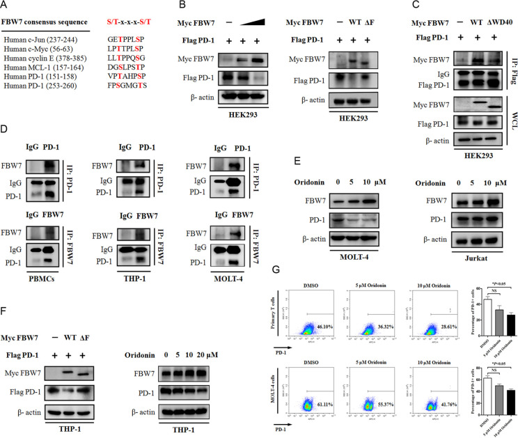
Methods: We used bioinformatic tools and biochemical methods to investigate the significance of F-box and WD repeat domain containing 7 (FBW7) in regulating PD-1 protein stability. By generating a panel of FBW7 and PD-1 encoding plasmids, we expressed FBW7 and PD-1 or their mutants to performed immunoprecipitation and immunoblotting assays. The efficacy of cotargeting FBW7 to enhance antitumor immunity was evaluated in C57BL/6J mice. These laboratory findings were further validated in tumor samples obtained from patients with non-small cell lung cancer (NSCLC).

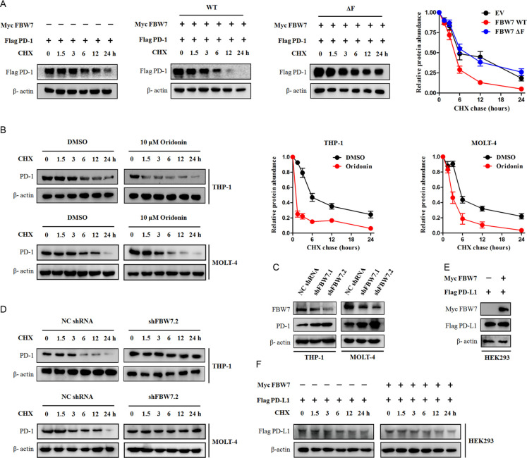
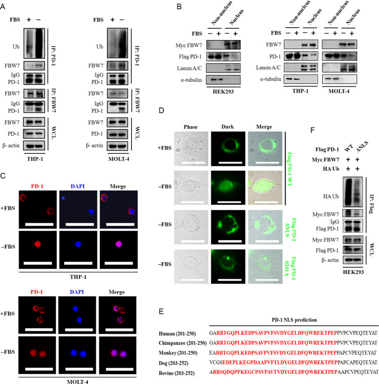
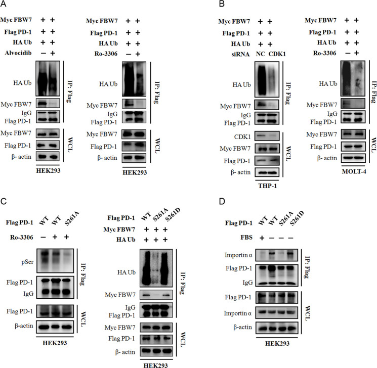
Results: We identified FBW7 as a E3 ubiquitin ligase for PD-1 protein, in which FBW7 promotes the K48-linked polyubiquitination of PD-1 protein at Lys233 residue. Cotargeting FBW7 accelerates PD-1 protein degradation and enhances antitumor immunity in vivo. Moreover, we demonstrated that cyclin-dependent kinase 1-mediated phosphorylation of Ser261 residue primes PD-1 protein nucleus translocation and binding with FBW7. Higher expression of FBW7 characterizes a 'hot' tumor microenvironment and confers more favorable responses to PD-1 blockade therapy.

Conclusions: This study highlights the critical role of FBW7 in determining PD-1 protein stability. FBW7 ubiquitinates PD-1 in a phosphorylation-dependent manner, as a consequence, leading to PD-1 protein degradation and cytotoxic lymphocytes infiltrating the tumor microenvironment. Screening FBW7 status would predict clinical response to anti-PD-1 immunotherapy in patients with NSCLC, and targeting FBW7 is a promising strategy to enhance antitumor immunity.

Keywords: Immunotherapy; Lung Neoplasms; Programmed Cell Death 1 Receptor; Translational Medical Research; Tumor Escape.


Conflict of interest statement

Competing interests: None declared.

Figures

Figure 1
Figure 1
FBW7 interacts with PD-1 protein. (A) Sequence alignment of the phosphodegron sequences recognized by FBW7 in c-Jun, c-Myc, cyclin E, MCL-1 and PD-1. Conserved degron sequences are shown in red. (B, C) Western blot analysis with antibodies specific for the indicated proteins of HEK293 cells transfected with Flag-tagged PD-1 and Myc-tagged FBW7 constructs. β-actin was used as equal loading control. (D) The primary PBMCs, THP-1 cells and MOLT-4 cells, which express endogenous PD-1 and FBW7, were precipitated with anti-PD-1/anti-FBW7 antibody or isotype IgG, and the resultant immunoprecipitates were subjected to immunoblotting. (E) The MOLT-4 and Jurkat cells were treated with increasing concentrations of oridonin and analyzed for PD-1 protein abundance by Western blot. (F) The THP-1 cells were transfected with indicated constructs or treated with increasing concentrations of oridonin. The expression of exogenous and endogenous FBW7 and PD-1 protein was determined by immunoblotting. (G) Primary T cells and MOLT-4 cells were treated with oridonin at final concentrations of 5 µM and 10 µM for 24 hours. After indicated treatment, cells were incubated with Alexa Fluor 647-conjugated anti-PD-1 antibody. Cell surface expression of PD-1 was assessed by flow cytometry. Representative flow cytometry images were shown. FBW7, F-box and WD repeat domain containing 7; PBMCs, peripheral blood mononuclear cells;DMSO, dimethyl sulfoxide.
Figure 2
Figure 2
FBW7 determines PD-1 protein stability. (A) HEK293 cells were transfected with Flag-tagged PD-1 together with indicated Myc-tagged FBW7 constructs. Twenty hours after transfection, cells were split into 6 cm dishes and treated with 25 µg/mL CHX. At the indicated time points, cell lysates were prepared and probed with the Flag-tag antibody. β-actin was used as equal loading control. (B) The THP-1 cells and MOLT-4 cells were treated with 10 µM oridonin or DMSO and evaluated for PD-1 protein half-time by CHX chase assay. (C) The THP-1 cells and MOLT-4 cells were infected with shRNA lentivirus against FBW7 or negative control (NC), and selected with puromycin to eliminate non-infected cells. The resultant cells were analyzed for FBW7 and PD-1 expression by Western blot. (D) The THP-1 cells and MOLT-4 cells stably expressing shRNA targeting FBW7 were treated with CHX and evaluated for PD-1 protein half-time. (E, F) The Flag-tagged PD-L1 and Myc-tagged FBW7 constructs were expressed in HEK293 cells and evaluated for PD-L1 protein half-time by CHX chase assay. CHX, cycloheximide; FBW7, F-box and WD repeat domain containing 7;DMSO, dimethyl sulfoxide.
Figure 3
Figure 3
FBW7 as a E3 uibiquitin ligase for PD-1 protein. (A) Western blot analysis of whole cell lysates (WCL) and immunoprecipitates derived from THP-1 and MOLT-4 cells treated with DMSO or oridonin (10 µM) for 24 hours, and cells were treated with 20 μM MG132 for additional 5 hours before harvesting. The ubiquitination status of endogenous PD-1 protein was determined by immunoprecipitation (IP). β-actin was used as equal loading control. (B) HEK293 cells were transfected with Flag PD-1 together with indicated Myc FBW7 construct. Thirty hours after transfection, cells were treated with 20 μM MG132 for additional 8 hours to block the proteasome pathway before cell collection. The cell lysates were subjected to IP and immunoblotting, respectively. (C) Immunoblotting analysis of Flag tag pull-down precipitates and WCL derived from HEK293 cells transfected with HA Ub, Myc FBW7 and indicated Flag PD-1 construct. Thirty hours after transfection, cells were treated with 20 μM MG132 for 8 hours before cell collection. (D) Myc-tagged FBW7, Flag-tagged PD-1, and His-tagged Ub WT, or Ub K48, or Ub K63 plasmids were expressed in HEK293 cells. PD-1 protein ubiquitination status was determined by Flag tag pull-down and anti-His immunoblotting. (E) Western blot analysis of WCL and immunoprecipitates derived from HEK293 cells transfected with Myc FBW7, HA Ub together with indicated Flag PD-1 construct. The ubiquitination status of Flag-tagged PD-1 protein was determined in the Flag tag pull-down precipitates by using anti-HA primary antibody. (F) The Flag-tagged PD-1 WT and its KR mutants were expressed in THP-1 cells. Thirty hours after transfection, cells were treated with or without MG132 before cell collection. The expression of Flag PD-1 protein was assessed by Western blot. FBW7, F-box and WD repeat domain containing 7; HA, hemagglutinin;DMSO, dimethyl sulfoxide.
Figure 4
Figure 4
The nucleus translocation of PD-1 protein is required for its interaction with FBW7. (A) Immunoblotting of WCL and immunoprecipitates from THP-1 and MOLT-4 cells treated with or without FBS for 6 hours. MG132 was added into the culture medium 5 hours before harvesting. The ubiquitination status of PD-1 was assessed by using an antibody against Ub. β-actin was used as equal loading control. (B) HEK293 cells were transfected with Myc-tagged FBW7 and Flg-tagged PD-1 constructs. The resultant cells, THP-1 and MOLT-4 cells were cultured with or without FBS for 6 hours. After indicated treatment, cell fraction assay was performed and the expression of FBW7 and PD-1 in the non-nucleus lysates and nucleus lysates was determined by immunoblotting. α-tubulin and Lamin A/C were used as equal loading control for non-nucleus protein and nucleus protein, respectively. (C) Representative immunofluorescent images of the PD-1 protein localization in THP-1 and MOLT-4 cells cultured with or without FBS. Scale bar=100 µm. (D) The EGFP-tagged PD-1 constructs were transiently expressed in HEK293 cells and the distribution of PD-1 protein was visualized under a fluorescent microscope. Representative fluorescent images were shown. Scale bar=20 µm. (E) Alignment analysis of putative NLS region of PD-1 protein across a panel species. The conserved NLS region is highlighted in red. (F) Immunoblotting with the anti-Flag tag precipitates and WCL derived from HEK293 cells expressing Myc FBW7, HA Ub and Flag PD-1 WT or Flag PD-1 ΔNLS constructs. MG132 was added for 8 hour before cell collection. FBW7, F-box and WD repeat domain containing 7; HA, hemagglutinin; NLS, nuclear localization signal; WCL, whole cell lysates; FBS, fetal bovine serum.
Figure 5
Figure 5
Phosphorylation of PD-1 prompts FBW7-mediated ubiquitination and protein destruction. (A) HEK293 cells were transfected with Myc FBW7, Flag PD-1 and HA Ub constructs. Thirty hours after transfection, cells were treated with the pan-CDK inhibitor Alvocidib (10 µM) or the selective CDK1 inhibitor Ro-3306 (10 µM), together with 20 μM MG132 for another 8 hours. The ubiquitination status and the binding with FBW7 were determined with anti-Flag immunoprecipitates. β-actin was used as equal loading control. (B) The THP-1 cells and MOLT-4 cells were transfected with indicated plasmids and treated with either siRNA targeting CDK1 or Ro-3306 (10 µM). The ubiquitination status of PD-1 and its binding with FBW7 were assessed as described. (C) The Flag-tagged PD-1 and its S261A/S261D mutant was expressed in HEK293 cells. Phosphorylation status of the serine residue was determined in the anti-Flag immunoprecipitates by using an anti-pSer antibody. The ubiquitination status of PD-1 and its binding with FBW7 were assessed as described. (D) The effect of Ser261 phosphorylation on PD-1 ubiquitination was evaluated in HEK293 cells. Cells were transfected with indicated plasmids and cultured in the presence or absence of FBS for 6 hours. The binding to nucleus cotranslocator importin α was assessed in the anti-Flag immunoprecipitates. FBW7, F-box and WD repeat domain containing 7; WCL, whole cell lysates;FBS, fetal bovine serum.
Figure 6
Figure 6
Cotargeting FBW7 enhances antitumor immunity in vivo. (A) The immune competent C57BL/6 J mice and the immune compromised nude mice were subcutaneously injected with LLC cells. The mice were treated with vehicle, oridonin, anti-PD-1 antibody or their combination. The tumor volume was routinely monitored. At the end of the experiment, the syngeneic tumor was carefully removed and photographed. NS, not significant; *p<0.05 vs vehicle. (B) Evaluation of cell apoptosis in the syngeneic tumor by TUNEL assay. Representative images were shown. Scale bar=100 µm. NS, not significant; *p<0.05 vs vehicle, **p<0.01 vs vehicle. (C) Representative images of IHC staining of Ki67, FBW7, PD-1 and CD8 in the syngeneic tumor. The enlarged window indicated the infiltration of CD8+CTLs in the tumor microenviroment. Scale bar=50 µm. (D) Measurement of hepatic and renal function in tumor bearing mice receiving indicated treatment. CTLs, cytotoxic lymphocytes; FBW7, F-box and WD repeat domain containing 7; LLC, Lewis lung cancer; AST, aspartate transaminase; ALT, alanine transaminase; BUN, blood urea nitrogen.
Figure 7
Figure 7
Screening FBW7 to predict response to anti-PD-1 immunotherapy in NSCLC. (A, B) A total number of 23 cases of surgical resected NSCLC were analyzed for FBW7 and PD-1 expression by IHC staining. Representative IHC images were shown. Scale bar=50 µm. Each NSCLC sample yielded an IHC H-score for FBW7 and PD-1, respectively. The correlation between FBW7 and PD-1 was determined by linear regression analysis.**p<0.01. (C) Evaluation of the tumor microenvironment in DCB (n=27) and NDB (n=21) patients. The panCK was used to distinguish cancer cells and non-cancerous cells. Representative IHC images for tumor nests (positive for panCK), FBW7, and infiltrating CD8+CTLs were shown. Scale bar=50 µm. ***p<0.001. CTLs, cytotoxic lymphocytes; DCB, durable clinical benefit; FBW7, F-box and WD repeat domain containing 7; NBD, no durable clinical benefit; NSCLC, non-small cell lung cancer; IHC, immunohistochemistry.
Figure 8
Figure 8
A schematic diagram of CDK1-mediated PD-1 and FBW7 interaction and their underlying implications for antitumor immunity. The PD-1 protein is primarily expressed in the cell membrane and FBW7 is expressed in the nucleus of immune cells. In the FBW7 OFF condition, PD-1 phosphorylation is suppressed by the CDK1 inhibitor and it could not undergo nucleus translocation. Thus, the PD-1 protein constitutively expresses in immune cells, which in return activates the PD-1/PD-L1 immune suppressive pathway and inhibits CTLs infiltration. Tumors harboring the FBW7low/PD-1high feature manifest as ‘cold tumor’ and are not sensitive to PD-1 blockade immunotherapy. In contrast, the CDK1-mediated phosphorylation of PD-1 protein at Ser261 residue triggers a rapid movement into the nucleus, where PD-1 binds to FBW7 and undergoes ubiquitination and destruction. Overexpression of FBW7 could enhance this interaction, as a consequence, leading to the suppression of PD-1/PD-L1 signaling (FBW7 ON). The CD8+CTLs infiltrates the FBW7high/PD-1low ‘hot tumor’, which confers increased responsiveness to anti-PD-1 immunotherapy. CTLs, cytotoxic lymphocytes; FBW7, F-box and WD repeat domain containing 7.

Similar articles

Cited by

References

    1. Salmaninejad A, Valilou SF, Shabgah AG, et al. . PD-1/PD-L1 pathway: basic biology and role in cancer immunotherapy. J Cell Physiol 2019;234:16824–37. 10.1002/jcp.28358 - DOI - PubMed
    1. Gong J, Chehrazi-Raffle A, Reddi S, et al. . Development of PD-1 and PD-L1 inhibitors as a form of cancer immunotherapy: a comprehensive review of registration trials and future considerations. J Immunother Cancer 2018;6:8. 10.1186/s40425-018-0316-z - DOI - PMC - PubMed
    1. Chen L, Han X. Anti-PD-1/PD-L1 therapy of human cancer: past, present, and future. J Clin Invest 2015;125:3384–91. 10.1172/JCI80011 - DOI - PMC - PubMed
    1. Peranzoni E, Lemoine J, Vimeux L, et al. . Macrophages impede CD8 T cells from reaching tumor cells and limit the efficacy of anti-PD-1 treatment. Proc Natl Acad Sci U S A 2018;115:E4041–50. 10.1073/pnas.1720948115 - DOI - PMC - PubMed
    1. Xin Yu J, Hodge JP, Oliva C, et al. . Trends in clinical development for PD-1/PD-L1 inhibitors. Nat Rev Drug Discov 2020;19:163–4. 10.1038/d41573-019-00182-w - DOI - PubMed

Publication types

MeSH terms