. 2022 Oct 15;209(8):1586-1594.
doi: 10.4049/jimmunol.2200309. Epub 2022 Sep 14.

Epitope Mapping of Therapeutic Antibodies Targeting Human LAG3

Affiliations

Epitope Mapping of Therapeutic Antibodies Targeting Human LAG3

Pragati Agnihotri et al. J Immunol. .

Abstract

Lymphocyte activation gene 3 protein (LAG3; CD223) is an inhibitory receptor that is highly upregulated on exhausted T cells in tumors and chronic viral infection. Consequently, LAG3 is now a major immunotherapeutic target for the treatment of cancer, and many mAbs against human (h) LAG3 (hLAG3) have been generated to block its inhibitory activity. However, little or no information is available on the epitopes they recognize. We selected a panel of seven therapeutic mAbs from the patent literature for detailed characterization. These mAbs were expressed as Fab or single-chain variable fragments and shown to bind hLAG3 with nanomolar affinities, as measured by biolayer interferometry. Using competitive binding assays, we found that the seven mAbs recognize four distinct epitopes on hLAG3. To localize the epitopes, we carried out epitope mapping using chimeras between hLAG3 and mouse LAG3. All seven mAbs are directed against the first Ig-like domain (D1) of hLAG3, despite their different origins. Three mAbs almost exclusively target a unique 30-residue loop of D1 that forms at least part of the putative binding site for MHC class II, whereas four mainly recognize D1 determinants outside this loop. However, because all the mAbs block binding of hLAG3 to MHC class II, each of the epitopes they recognize must at least partially overlap the MHC class II binding site.


Figures

Figure 1.
Figure 1.
Purification of Fabs and Fab–hLAG3 and scFv–hLAG3 complexes. (A) Reducing 16% SDS-PAGE gel of purified recombinant Fab and scFv fragments used for affinity measurements and epitope binning (Coomassie blue G-250 staining). Lane 1, markers; lane 2, Fab 496G6; lane 3, Fab 4A10; lane 4, Fab 22D2; lane 5, Fab BAP050; lane 6, Fab 11C9; lane 7, Fab 13E2; lane 8, scFv relatlimab. (B) Superdex 200 size exclusion chromatography profiles of Fabs and scFv following streptavidin affinity chromatography. The column was previously calibrated with thyroglobulin, ferritin, aldolase, conalbumin, and ovalbumin as molecular mass standards. (C) Reducing 16% SDS-PAGE gel of purified Fab–hLAG3 and scFv–hLAG3 complexes. The complexes were prepared by co-transfecting vectors encoding hLAG3 and Fab or scFv fragments into Expi293 cells. Lane 1, markers; lane 2, Fab 496G6–hLAG3; lane 3, Fab 4A10–hLAG3; lane 4, Fab 22D2–hLAG3; lane 5, Fab BAP050–hLAG3; lane 6, Fab 11C9–hLAG3; lane 7, Fab 13E2–hLAG3; lane 8, scFv relatlimab–hLAG3. (D) Superdex 200 size exclusion chromatography profiles of Fab–hLAG3 and scFv–hLAG3 complexes following streptavidin affinity chromatography.
Figure 2.
Figure 2.
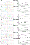
BLI analysis of Fab and scFv binding to hLAG3. (A) Sensograms (left) for Fab 4A10 binding to immobilized hLAG3. Fab 4A10 concentrations were 100, 50, 25, 12.5, 6.25, and 3.15 nM. Steady-state analysis graph (right) gave a KD of 41 ± 4.8 nM. (B) Sensograms (left) for Fab 496G6 binding to immobilized hLAG3. Steady-state analysis graph (right) gave a KD of 13 ± 1.8 nM. (C) Sensograms (left) for Fab 22D2 binding to immobilized hLAG3. Steady-state analysis graph (right) gave a KD of 5.0 ± 1.7 nM. (D) Sensograms (left) for Fab BAP050 binding to immobilized hLAG3. Steady-state analysis graph (right) gave a KD of 9.0 ± 0.07 nM. (E) Sensograms (left) for Fab 11C9 binding to immobilized hLAG3. Steady-state analysis graph (right) gave a KD of 11 ± 0.06 nM. (F) Sensograms (left) for Fab 13E2 binding to immobilized hLAG3. Steady-state analysis graph (right) gave a KD of 2.3 ± 0.1 nM. (G) Sensograms (left) for scFv relatlimab binding to immobilized hLAG3. Steady-state analysis graph (right) gave a KD of 1.8 ± 0.18 nM.
Figure 3.
Figure 3.
Epitope binning of anti-LAG3 antibodies by BLI. Binning experiments were performed with His6-tagged hLAG3 immobilized on Ni2+-NTA biosensors. The binding step with saturating antibody followed by binding/blocking antibodies is presented for each indicated saturating antibody. In each set of experiments, the background signal was obtained from a parallel sensor incubated with the saturating antibody as control. (A) Sensograms with 4A10 as saturating antibody. (B) Sensograms with 496G6 as saturating antibody. (C) Sensograms with 22D2 as saturating antibody. (D) Sensograms with BAP050 as saturating antibody. (E) Sensograms with 11C9 as saturating antibody. (F) Sensograms with 13E2 as saturating antibody. (G) Sensograms with relatlimab as saturating antibody. (H) Matrix with relative binding values calculated by Octet Analysis Software 12.0. Saturating antibodies are listed in the left column and blocking antibodies in the top row. (I) Four noncompeting hLAG3 binding epitopes were identified and classified into four groups: I (496G6, 22D2, and 13E2); II (4A10 and relatlimab); III (BAP050 and 496G6); and IV (11C9 and BAP050).
Figure 4.
Figure 4.
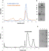
Preparation of Fab2–hLAG3 triple complexes. (A) Superdex 200 size exclusion chromatography profiles of Fab 22D2–Fab 11C9–hLAG3 and Fab 22D2–Fab BAP050–hLAG3 complexes following Ni2+-NTA affinity chromatography. (B) Reducing 16% SDS-PAGE gel of purified Fab2–hLAG3 complexes. Lane 1, Fab 22D2–Fab 11C9–hLAG3; lane 2, markers; lane 3, Fab 22D2–Fab BAP050–hLAG3. (C) Superdex 200 profile of mixture of purified Fab 22D2–hLAG3 complex and purified Fab 13E2. The vertical arrow indicates expected position of an Fab2–hLAG3 triple complex. (D) Reducing 16% SDS-PAGE of mixture of Fab 22D2–hLAG3 and Fab 13E2 that was loaded on Superdex 200 column (lane 1), major peak in size exclusion profile (lane 2), Fab 13E2 (lane 3), and markers (lane 4).
Figure 5.
Figure 5.
Epitope mapping of anti-hLAG3 antibodies. 293T cells were transfected with plasmids containing deletions and domain swap mutants of hLAG3 and mLAG3, stained with the panel of anti-hLAG3 mAbs, and analyzed by flow cytometry. (A) Schematic diagrams on left depict deletion and domain-swapped mutants of hLAG3 and mLAG3. Heat map on right shows binding of mAbs to the mutants. Numbers indicate fold-increase of mean fluorescence intensity over vector only and are averages of 6 experiments ± SE. (B) Comparison of the amino acid sequences of D1 loop region of hLAG3 and mLAG3 with conserved residues highlighted in red.

Similar articles

Cited by

References

    1. Topalian SL, Drake CG, and Pardoll DM. 2015. Immune checkpoint blockade: a common denominator approach to cancer therapy. Cancer Cell 13: 450–461. - PMC - PubMed
    1. Driessens G, Kline J, and Gajewski TF. 2009. Costimulatory and coinhibitory receptors in anti-tumor immunity. Immunol. Rev 229: 126–144. - PMC - PubMed
    1. Hodi FS, O'Day SJ, McDermott DF, Weber RW, Sosman JA, Haanen JB, Gonzalez R, Robert C, Schadendorf D, Hassel JC, et al. 2010. Improved survival with ipilimumab in patients with metastatic melanoma. N. Engl. J. Med 363: 711–723. - PMC - PubMed
    1. Topalian SL, Sznol M, McDermott DF, Kluger HM, Carvajal RD, Sharfman WH, Brahmer JR, Lawrence DP, Atkins MB, Powderly JD, et al. 2014. Survival, durable tumor remission, and long-term safety in patients with advanced melanoma receiving nivolumab. J. Clin. Oncol 32: 1020–1030. - PMC - PubMed
    1. Larkin J, Chiarion-Sileni V, Gonzalez R, Grob JJ, Rutkowski P, Lao CD, Cowey CL, Schadendorf D, Wagstaff J, Dummer R, et al. 2019. Five-year survival with combined nivolumab and ipilimumab in advanced melanoma. N. Engl. J. Med 381: 1535–1546. - PubMed

Publication types