

Usefulness of Ultrasonography in the Diagnosis and Follow-up of Extracranial Vertebral Artery Dissection
- PMID: 36104189
- PMCID: PMC10183284
- DOI: 10.2169/internalmedicine.0019-22
Usefulness of Ultrasonography in the Diagnosis and Follow-up of Extracranial Vertebral Artery Dissection
Abstract
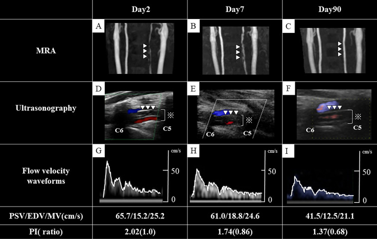
Extracranial vertebral artery dissection is a cerebrovascular disease that occurs most commonly in young people. A 32-year-old man experienced sudden cervical pain and was diagnosed with left vertebral artery dissection after arterial changes were identified by ultrasonography. The reduction in the size of an intramural hematoma in the left vertebral artery and in the peak systolic velocity were evaluated over time. Computed tomography, magnetic resonance imaging, and cerebral angiography are generally performed to diagnose and follow-up extracranial vertebral artery dissection; however, carotid ultrasonography has an advantage over these modalities by enabling the simultaneous observation of vascular morphology and hemodynamics.
Keywords: ultrasonography; vertebral artery dissection.
Conflict of interest statement
Figures
References
-
- Bejor Y, Daubail B, Debette S, et al. . Incidence and outcome of cerebrovascular events related to cervical artery dissection; the Dijon Stroke Registry. Int J Stroke 9: 879-882, 2014. - PubMed
-
- Kwon JY, Kim NY, Suh DC, et al. . Intracranial and extracranial artery dissection presenting with ischemic stroke: lesion location and stroke mechanism. J Neurol Sci 358: 371-376, 2015. - PubMed
-
- Provenzale JM, Sarikaya B. Comparison of test performance characterisics of MRI, MR angiography, and CT angiography in the diagnosis of carotid and vertebral artery dissection: a review of the medical literature. AJR Am J Roentgenol 193: 1167-1174, 2009. - PubMed
-
- Markus HS, Hayter E, Levi C, et al. . Antiplatelet treatment compared with anticoagulation treatment for cervical artery dissection (CADISS): a randomized trial. Lancet Neurol 14: 361-367, 2015. - PubMed