. 2022 Sep 14;12(1):15453.
doi: 10.1038/s41598-022-19113-8.

Cerebrovascular reactivity in multiple sclerosis is restored with reduced inflammation during immunomodulation

Affiliations

Cerebrovascular reactivity in multiple sclerosis is restored with reduced inflammation during immunomodulation

Antonio Maria Chiarelli et al. Sci Rep. .

Abstract

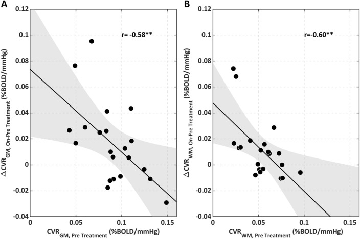
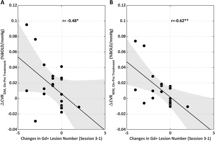
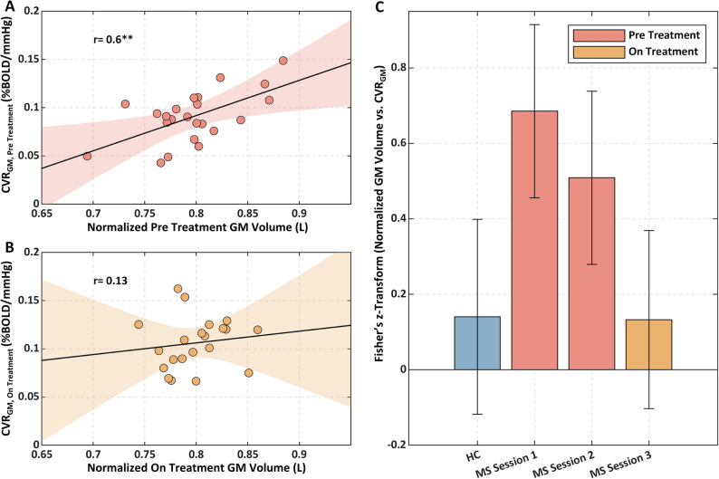
Cerebrovascular reactivity (CVR) reflects the capacity of the brain's vasculature to increase blood flow following a vasodilatory stimulus. Reactivity is an essential property of the brain's blood vessels that maintains nutrient supplies in the face of changing demand. In Multiple Sclerosis (MS), CVR may be diminished with brain inflammation and this may contribute to neurodegeneration. We test the hypothesis that CVR is altered with MS neuroinflammation and that it is restored when inflammation is reduced. Using a breath-hold task during functional Magnetic Resonance Imaging (MRI), we mapped grey matter and white matter CVRs (CVRGM and CVRWM, respectively) in 23 young MS patients, eligible for disease modifying therapy, before and during Interferon beta treatment. Inflammatory activity was inferred from the presence of Gadolinium enhancing lesions at MRI. Eighteen age and gender-matched healthy controls (HC) were also assessed. Enhancing lesions were observed in 12 patients at the start of the study and in 3 patients during treatment. Patients had lower pre-treatment CVRGM (p = 0.04) and CVRWM (p = 0.02) compared to HC. In patients, a lower pre-treatment CVRGM was associated with a lower GM volume (r = 0.60, p = 0.003). On-treatment, there was an increase in CVRGM (p = 0.02) and CVRWM (p = 0.03) that negatively correlated with pre-treatment CVR (GM: r = - 0.58, p = 0.005; WM: r = - 0.60, p = 0.003). CVR increased when enhancing lesions reduced in number (GM: r = - 0.48, p = 0.02, WM: r = - 0.62, p = 0.003). Resolution of inflammation may restore altered cerebrovascular function limiting neurodegeneration in MS. Imaging of cerebrovascular function may thereby inform tissue physiology and improve treatment monitoring.


Conflict of interest statement

Valentina Tomassini received funds for honoraria, research support and travel grants from Biogen, Merck Serono, Roche, Novartis, Bristol Myers Squibb, Viatris, Almirall, Sanofi. The other Authors declare no competing financial and non-financial interests.

Figures

Figure 1
Figure 1
Timeline of the study together with eligibility criteria and MRI sequences performed.
Figure 2
Figure 2
(A) Top: Example of raw CO2 and PetCO2 traces during the breath-hold (BH) task (BH periods highlighted in yellow). Middle: PetCO2 trace after resampling, quadratic detrending and convolution with the cHRF. Bottom: Processed PetCO2 trace overlaid onto average GM and WM fractional BOLD (%). Processed PetCO2 trace was used as a regressor for the voxel-wise GLM analysis. (B) Top: Example of brain tissue type segmentation in GM, WM and CSF in one exemplar subject. Middle: Unsmoothed CVR map obtained within the GLM framework in the same subject. Bottom: Voxel-wise distributions of CVR within GM and WM. The median values of such distributions were considered as global estimates of CVRs within the two tissue compartments.
Figure 3
Figure 3
Global average ± SD of the mean CVRs for HC and MS in the three sessions. (A) CVR in GM; on the left, mean and associated standard error; on the right, violin plots representing the sample distribution of CVR. (B) CVR in WM; on the left, mean and associated standard error; on the right, violin plots. *p < 0.05.
Figure 4
Figure 4
On vs. pre-treatment changes in CVR as a function of the pre-treatment CVR (A) in the GM and (B) in the WM. **p < 0.01.
Figure 5
Figure 5
Maps of on vs. pre-treatment change in CVR in MS patients: (A) Map in absolute units; (B) Statistical Parametric (t-score) map; (C) thresholded (p < 0.05) map of null-hypothesis probability, corrected for multiple comparison using the threshold free cluster enhancement (TFCE) approach. Coordinates (in mm) refers to the MNI152 template.
Figure 6
Figure 6
On vs. pre-treatment changes in CVR as a function of the changes in the number of lesions between session 3 and session 1 (A) in the GM and (B) in the WM. *p < 0.05, **p < 0.01.
Figure 7
Figure 7
Scatterplots of normalised GM volume vs. CVR in GM for MS (A) pre-treatment and (B) on-treatment; (C) Fisher’s z-transforms of the correlations between normalised tissue volumes with CVR in GM and WM for HC and MS patients in the three sessions. **p < 0.01.

References

    1. D’haeseleer M, Hostenbach S, Peeters I, et al. Cerebral hypoperfusion: A new pathophysiologic concept in multiple sclerosis? J. Cereb. Blood Flow Metab. 2015;35:1406–1410. doi: 10.1038/jcbfm.2015.131. - DOI - PMC - PubMed
    1. Frischer JM, Bramow S, Dal-Bianco A, et al. The relation between inflammation and neurodegeneration in multiple sclerosis brains. Brain. 2009;132:1175–1189. doi: 10.1093/brain/awp070. - DOI - PMC - PubMed
    1. Trapp BD, Ransohoff RM, Fisher E, Rudick RA. Neurodegeneration in multiple sclerosis: Relationship to neurological disability. Neuroscientist. 1999;5:48–57. doi: 10.1177/107385849900500107. - DOI
    1. Paling D, Thade Petersen E, Tozer DJ, et al. Cerebral arterial bolus arrival time is prolonged in multiple sclerosis and associated with disability. J. Cereb. Blood Flow Metab. 2014;34:34–42. doi: 10.1038/jcbfm.2013.161. - DOI - PMC - PubMed
    1. Rashid W, Parkes LM, Ingle GT, et al. Abnormalities of cerebral perfusion in multiple sclerosis. J. Neurol. Neurosurg. Psychiatry. 2004;75:1288–1293. doi: 10.1136/jnnp.2003.026021. - DOI - PMC - PubMed

Publication types