. 2022 Jun;15(6):1389-1404.
doi: 10.1038/s41385-022-00566-z. Epub 2022 Sep 14.

A lentiviral vector expressing a dendritic cell-targeting multimer induces mucosal anti-mycobacterial CD4+ T-cell immunity

Affiliations

A lentiviral vector expressing a dendritic cell-targeting multimer induces mucosal anti-mycobacterial CD4+ T-cell immunity

François Anna et al. Mucosal Immunol. 2022 Jun.

Abstract

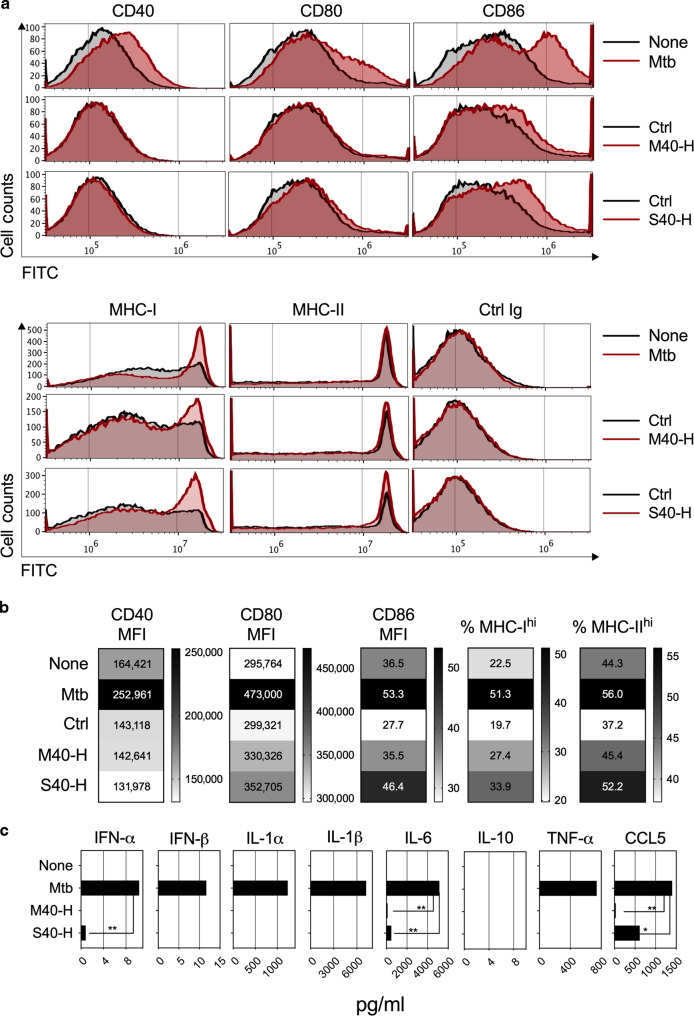
Most viral vectors, including the potently immunogenic lentiviral vectors (LVs), only poorly direct antigens to the MHC-II endosomal pathway and elicit CD4+ T cells. We developed a new generation of LVs encoding antigen-bearing monomers of collectins substituted at their C-terminal domain with the CD40 ligand ectodomain to target and activate antigen-presenting cells. Host cells transduced with such optimized LVs secreted soluble collectin-antigen polymers with the potential to be endocytosed in vivo and reach the MHC-II pathway. In the murine tuberculosis model, such LVs induced efficient MHC-II antigenic presentation and triggered both CD8+ and CD4+ T cells at the systemic and mucosal levels. They also conferred a significant booster effect, consistent with the importance of CD4+ T cells for protection against Mycobacterium tuberculosis. Given the pivotal role of CD4+ T cells in orchestrating innate and adaptive immunity, this strategy could have a broad range of applications in the vaccinology field.


Conflict of interest statement

P.C. is the founder and CSO of TheraVectys. F.A., F.M., P.A., A.N., and F.N. are employees of TheraVectys. F.A., J.L., F.M., C.B., P.C., and L.M. are inventors of a pending patent directed to the optimized vaccination LV, able to induce CD4+ T-cell responses. L.M. has a consultancy activity for TheraVectys. Other authors declare no competing interests.

Figures

Fig. 1
Fig. 1. Structure of MBL- and SPD-based antigen carriers.
Schematic structure of the MBL and SPD collectin polymers. a Structural domains of MBL and SPD. CRD = carbohydrate-recognition domain. b MBL and SPD self-assembled collagen-like triple helixes formed by interchain cysteine bonds. c SPD cross-shaped dodecamer. d SPD and MBL “tulip-bouquet” octodecamers, adapted from. ef Schematic representation of the primary structure of the designed M40 (e) and S40 (f) monomers carrying selected Mtb antigens: crosslinking region (S), collagen-like region (Coll), neck region (N).
Fig. 2
Fig. 2. Multimerization of S40-based carriers.
a Western-blot analysis of supernatants and cell lysates of HEK293T cells, either transduced at an MOI = 100 of LV::S40-H-StrepII or non-transduced, in reducing or non-reducing conditions. β-Actin staining was use of as loading control. b Chromatogram of column calibration using a set of purified recombinant proteins on a Superose 6 FPLC column. X axis correspond to MW equivalence observed during elution and Y axis to UV absorbance (280 nm). c Chromatogram of 50x-concentrated supernatant of HEK293T cells transduced with LV::S40-H-StrepII. d Quantification of S40-H-StrepII protein by measuring mean pixel intensity of the band of each well, corresponding to the molecular weight of monomeric S40-H-StrepII. e Western blot analysis of elution fractions in reducing conditions.
Fig. 3
Fig. 3. Properties of backbones at recruiting APC and inducing antigenic presentation by both the MHC-I and-II pathways.
a, c BM-DCs from BALB/c (H-2d) or C57BL/6 (H-2b) mice were transduced (MOI = 20) with LV::M40-H, -HA, -HAP, or -HAPE (a) or LV::S40-H, -HAPEHR, or -HAPEHR-20 (c) under the transcriptional control of the BCUAG promoter. Control cells were transduced with LV::EsxH alone. b, d BM-DCs from BALB/c or C57BL/6 mice were incubated with successive dilutions of supernatants of HEK-293T cells transduced (MOI = 20) for 48 h with each of the indicated LVs. On day 3 after addition of the LVs or day 1 after incubation with the HEK293T cell supernatants, the presentation of MHC-I- or -II-restricted epitopes of the EsxH, EsxA, PE19, or EspC mycobacterial antigens by DCs was assessed by their co-culture with T-cell hybridomas specific for EsxH:20-28 (YB8 cell line, restricted by Kd), EsxH:74-88 (1G1 cell line, restricted by I-Ad), EsxA:1-20 (NB11 cell line, restricted by I-Ab), PE:19:1-18 (IF6 cell line, restricted by I-Ab), or EspC:45:54 (IF1 cell line, restricted by I-Ab). Results are presented as the concentration of IL-2 produced by the T-cell hybridomas 24 h after the beginning of the co-cultures. The amount of IL-2 found in the co-culture supernatants is proportional to the efficacy of antigenic presentation by DCs and TCR triggering. This assay does not measure the physiological response induced in the T cells but only provides an indicator of the stimulation of the T-cell hybridoma TCR. e Impact of HAPEHR and HAPEHR-20 proteins on migration of BM-DCs in a transwell system. Supernatant of HEK293T cells transduced (MOI = 100) with LV::S40-HAPEHR or LV::S40-HAPEHR-20 (n = 6). Statistical significance was evaluated using the Mann–Whitney test (* = p < 0.05, ** = p < 0.01, *** = p < 0.001, ns non-significant).
Fig. 4
Fig. 4. Phenotypic Maturation of DCs Induced by M40 or S40.
a Phenotypic maturation of BM-DCs from C57BL/6 mice infected at an MOI of 5 with Mtb, as a positive control, or incubated with supernatants from HEK-293T cells transduced (MOI = 20) with LV::EsxH alone (Ctrl), LV::M40-H, or LV::S40-H. Expression of co-stimulatory or MHC molecules on the surface of CD11b+ CD11c+ cells at 24 h post-immunization was assessed by cytometry. b Heatmaps showing the mean fluorescence intensity (MFI) of CD40 and CD80 surface expression and the percentage of CD86hi, MHC-I hi, and MHC-II hi DCs. c Quantitation of inflammatory mediators in the culture supernatants of the same DCs. Results are representative of at least two independent experiments.
Fig. 5
Fig. 5. Immunogenicity of Multi-Antigenic LV::S40-HAPEHR or LV::S40-HAPEHR-20.
a IFN-γ T-cell responses, as assessed by ELISPOT on day 13 post-immunization, in the spleens of individual C57BL/6 mice (n = 3) immunized s.c. with 1 × 108 TU/mouse of LV::S40-HAPEHR or LV::S40-HAPEHR-20. The frequency of responding T cells was determined following in vitro stimulation with the indicated synthetic peptides. Quantitative differences between the groups of mice immunized with LV::S40-HAPEHR or LV::S40-HAPEHR-20, were not statistically significant (Mann–Whitney test). b Epitope mapping of Hrp-1 and RfpD, as determined using pooled splenocytes from 3 C57BL/6 mice/group injected with PBS or immunized s.c. with 1 × 108 TU/mouse of LV::S40-HAPEHR prior to stimulation with each of the individual peptides from the Hrp-1- or RpfD-derived overlapping 15-mers offset by five amino acids. c Cytometric analysis of intracellular IFN-γ vs IL-2 staining of CD4+ T splenocytes after stimulation with 10 µg/ml of the indicated peptides encompassing the immunodominant epitopes identified in b. Pooled splenocytes from three mice/group were used.
Fig. 6
Fig. 6. Features of mucosal CD4+ T cells triggered by i.n. immunization with LV::S40-HAPEHR or LV::S40-HAPEHR-20.
C57BL/6 mice (n = 3/group) were immunized i.n. with 1 × 108 TU of LV::S40-HAPEHR or LV::S40-HAPEHR-20. On 14 dpi, lung CD4+ T cells were distinguished by their location within the interstitium (CD45i.v) or vasculature (CD45i.v+) by an i.v. injection of PE-anti-CD45 mAb. a Profile of CD27 vs CD62L or CD45RB, and b CD103 vs CD69, or CD44 vs CXCR3 of lung CD4+ T cells of the interstitium or vasculature. c Percentage of (poly)functional CD4+ T cells specific to EsxA, PE19, or EspC in the lung interstitium or vasculature, as determined by ICS. Results, representative of two independent experiments, were obtained using pooled lungs for each group (n = 3/group). Individual points represent technical duplicates. Results are representative of at least three independent experiments.
Fig. 7
Fig. 7. Features of mucosal CD8+ T cells induced by i.n. immunization with LV::S40-HAPEHR or LV::S40-HAPEHR-20.
The immunized C57BL/6 mice are those studied in the Fig. 6. a Shown are lung CD8+ T cells, distinguished for their location within the interstitium (CD45i.v) or in the vasculature (CD45i.v+). Profile of CD27 vs CD62L or CD45RB, and b CD103 vs CD69, or CD44 vs CXCR3 of the lung CD8+ T cells from the interstitium or vasculature. c Recapitulative percentages of (poly)functional CD8+ T cells specific to EsxH or EspC in the lung interstitium (top) or vasculature (bottom), as determined by ICS. Results are representative of at least two independent experiments.
Fig. 8
Fig. 8. Protective potential of an optimized poly-antigenic LV as a booster against Mtb.
a Timeline of the prime-boost-challenge performed in C57BL/6 mice (n = 5–8 mice/group). b Mtb burden as quantitated by CFU counting in the lungs of BCG::ESX-1Mmar-primed and LV::S40-HAPEHR-20-boosted mice on week 5 post challenge. c Whole-lung section of the left lobe and d quantification of the number and size of lung granulomatous lesions per mouse in each experimental group. e Mtb burden quantified by CFU counting in the lungs of BCG-primed and LV::S40-HAPEHR-20-boosted C57BL/6 mice following the timeline indicated in a, but using Danish BCG for the prime immunization and i.n. Mtb challenge. The pooled results from two independent experiments are shown (n = 6–8 mice/group in each experiment). The significance of the differences was determined using the Mann–Whitney test. ns not significant.

References

    1. Di Nunzio F, et al. HIV-derived vectors for therapy and vaccination against HIV. Vaccine. 2012;30:2499–2509. doi: 10.1016/j.vaccine.2012.01.089. - DOI - PubMed
    1. Hu B, Tai A, Wang P. Immunization delivered by lentiviral vectors for cancer and infectious diseases. Immunol. Rev. 2011;239:45–61. doi: 10.1111/j.1600-065X.2010.00967.x. - DOI - PMC - PubMed
    1. Rowe HM, et al. Immunization with a lentiviral vector stimulates both CD4 and CD8 T cell responses to an ovalbumin transgene. Mol. Ther. 2006;13:310–319. doi: 10.1016/j.ymthe.2005.08.025. - DOI - PubMed
    1. Thiele F, et al. Modified vaccinia virus Ankara-infected dendritic cells present CD4+ T-cell epitopes by endogenous major histocompatibility complex class II presentation pathways. J. Virol. 2015;89:2698–2709. doi: 10.1128/JVI.03244-14. - DOI - PMC - PubMed
    1. Zhu J, Paul WE. Heterogeneity and plasticity of T helper cells. Cell Res. 2010;20:4–12. doi: 10.1038/cr.2009.138. - DOI - PMC - PubMed

Publication types

LinkOut - more resources