

Foreign body-type giant cell reaction with extensive granulation tissue and intense inflammation after chemotherapy mimicking residual lymphoma on FDG PET
- PMID: 36104639
- PMCID: PMC9474748
- DOI: 10.1186/s41824-022-00137-2
Foreign body-type giant cell reaction with extensive granulation tissue and intense inflammation after chemotherapy mimicking residual lymphoma on FDG PET
Abstract
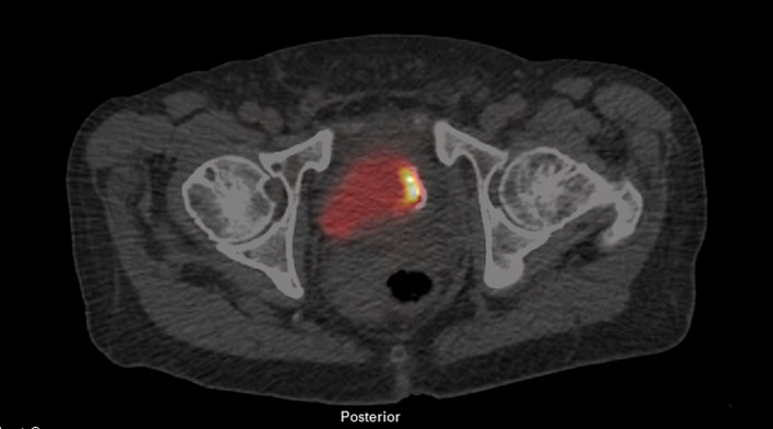
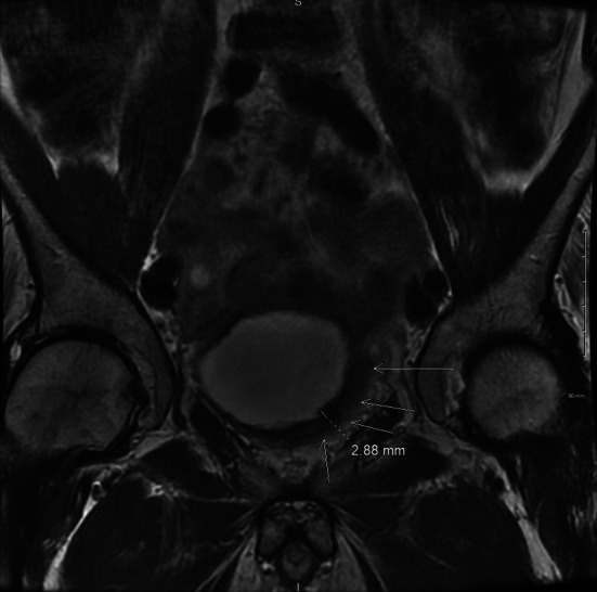
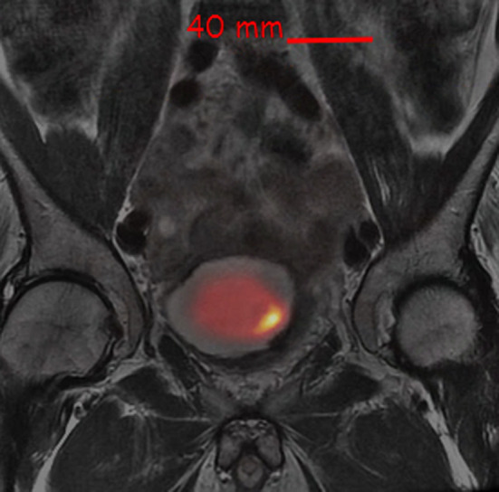
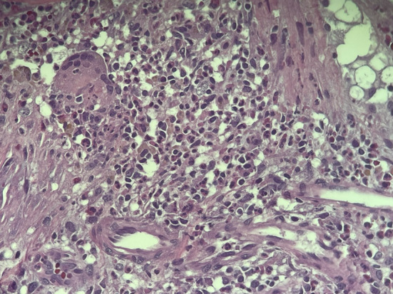
Foreign body-type giant cell reaction is typically a biological and immunological reaction to the presence of foreign bodies such as catheters, parasites or biomaterials with a collection of fused macrophages (giant cell). We reported an unusual case of [18F]FDG PET findings in diffuse large B cell lymphoma in the urinary bladder following incomplete resection and chemotherapy. As the restaging [18F]FDG PET showed intense [18F]FDG uptake in the urinary bladder at the resection site concerning for recurrence, the lesion was subsequently resected and histopathology showed extensive granulation tissue with foreign body-type giant cell reaction with no suspected foreign bodies or neoplasia.
Keywords: Diffuse large B cell lymphoma; Foreign body-type giant cell reaction; [18F]FDG PET.
© 2022. The Author(s).
Conflict of interest statement
The authors declare that there are no competing interests.
Figures
Similar articles
-
More advantages in detecting bone and soft tissue metastases from prostate cancer using 18F-PSMA PET/CT.Hell J Nucl Med. 2019 Jan-Apr;22(1):6-9. doi: 10.1967/s002449910952. Epub 2019 Mar 7. Hell J Nucl Med. 2019. PMID: 30843003
-
All That Glitters Is Not Gold" - A Case of an Occult Foreign Body in the Lung with Elevated 2-[18F]-Fluoro-2-deoxy-D-glucose (FDG) Uptake Mimicking Bronchogenic Carcinoma.Cureus. 2017 Jan 23;9(1):e990. doi: 10.7759/cureus.990. Cureus. 2017. PMID: 28265526 Free PMC article.
-
18 F-FDG Whole-Body PET-MR in Primary Hepatic Lymphoma Mimicking Focal Nodular Hyperplasia.World J Nucl Med. 2022 Sep 9;21(4):338-341. doi: 10.1055/s-0042-1750398. eCollection 2022 Dec. World J Nucl Med. 2022. PMID: 36398300 Free PMC article.
-
An update on the role of interim restaging FDG-PET in patients with diffuse large B-cell lymphoma and Hodgkin lymphoma.J Natl Compr Canc Netw. 2010 Mar;8(3):347-52. doi: 10.6004/jnccn.2010.0023. J Natl Compr Canc Netw. 2010. PMID: 20202464 Review.
-
18F-FDG PET/CT in the diagnosis of an extranodal relapse of diffuse large B-cell lymphoma (DLBCL): a clinical case with a literature review.Nucl Med Rev Cent East Eur. 2016;19(B):11-13. doi: 10.5603/NMR.2016.0029. Nucl Med Rev Cent East Eur. 2016. PMID: 27813622 Review.
References
LinkOut - more resources
Full Text Sources
Research Materials