. 2022 Sep 15;20(1):315.
doi: 10.1186/s12916-022-02500-3.

Single-cell analysis of menstrual endometrial tissues defines phenotypes associated with endometriosis

Affiliations

Single-cell analysis of menstrual endometrial tissues defines phenotypes associated with endometriosis

Andrew J Shih et al. BMC Med. .

Abstract

Background: Endometriosis is a common, complex disorder which is underrecognized and subject to prolonged delays in diagnosis. It is accompanied by significant changes in the eutopic endometrial lining.

Methods: We have undertaken the first single-cell RNA-sequencing (scRNA-Seq) comparison of endometrial tissues in freshly collected menstrual effluent (ME) from 33 subjects, including confirmed endometriosis patients (cases) and controls as well as symptomatic subjects (who have chronic symptoms suggestive of endometriosis but have not been diagnosed).

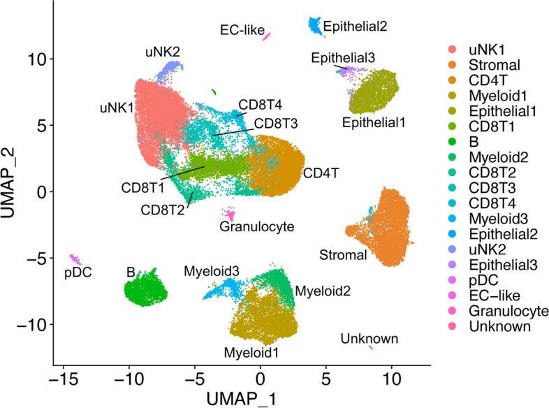
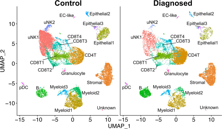
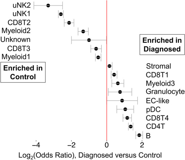
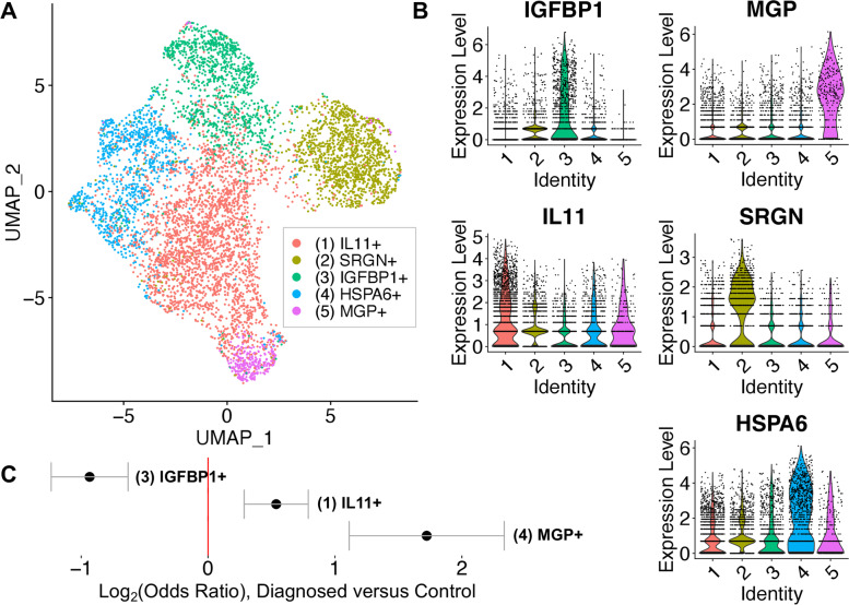
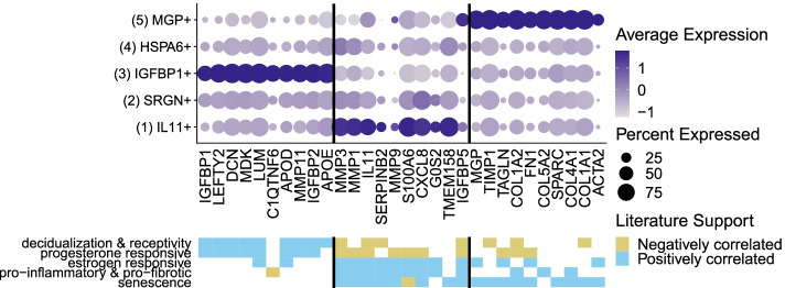
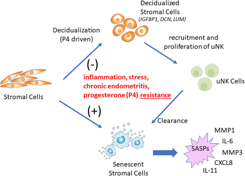
Results: We identify a unique subcluster of proliferating uterine natural killer (uNK) cells in ME-tissues from controls that is almost absent from endometriosis cases, along with a striking reduction of total uNK cells in the ME of cases (p < 10-16). In addition, an IGFBP1+ decidualized subset of endometrial stromal cells are abundant in the shed endometrium of controls when compared to cases (p < 10-16) confirming findings of compromised decidualization of cultured stromal cells from cases. By contrast, endometrial stromal cells from cases are enriched in cells expressing pro-inflammatory and senescent phenotypes. An enrichment of B cells in the cases (p = 5.8 × 10⁻⁶) raises the possibility that some may have chronic endometritis, a disorder which predisposes to endometriosis.

Conclusions: We propose that characterization of endometrial tissues in ME will provide an effective screening tool for identifying endometriosis in patients with chronic symptoms suggestive of this disorder. This constitutes a major advance, since delayed diagnosis for many years is a major clinical problem in the evaluation of these patients. Comprehensive analysis of ME is expected to lead to new diagnostic and therapeutic approaches to endometriosis and other associated reproductive disorders such as female infertility.

Keywords: Biomarkers; Decidualization; Endometriosis; Fibrosis; Inflammation; Menstrual blood; Menstrual effluent; Senescence; Single-cell RNA sequencing.


Conflict of interest statement

The authors declare that they have no competing interests.

Figures

Fig. 1
Fig. 1
ME contains endometrial tissues. Histological analysis of endometrial tissues isolated from the menstrual effluent (ME) from 4 separate subjects: A control subject, B, C two subjects with pathologically confirmed endometriosis, and D subject with chronic symptoms of endometriosis (not yet diagnosed). Upper panels for AD: H&E staining is shown in two panels at two magnifications for each individual: A 40X (left) and 200X (right), B 100X and 200X, C 100X and 200X, and D 40X and 200X; arrowheads point to glandular epithelium. Sections show typical late secretory/menstrual endometrium with expanded stroma containing scattered inflammatory cells and secretory and inactive type glands. Lower panels for AD: immunostaining with anti-CD10 and anti-CD56 antibodies to detect stromal cells (left) and uterine NK (uNK) cells (right), respectively, at 100X. Scale bars are shown in each image
Fig. 2
Fig. 2
Cellular composition of digested ME based on scRNA-Seq. UMAP plot for all 33 digested menstrual effluent (ME) samples (controls = 9; endometriosis cases = 11; symptomatic cases = 13). Several well-delineated cell clusters include a large cluster of uterine NK cells (uNK1), as well as clearly separated stromal cells, epithelial cells, and B cells. Several clusters each of T cells and myeloid cells are also defined, as well as a small cluster of plasmacytoid dendritic cells (pDC). A small cluster of approximately 60 unknown cells is in the lower right corner. The positive gene markers used to generate the cell clusters shown are included in Additional file 2.
Fig. 3
Fig. 3
Distinct cellular composition differences in digested ME from endometriosis cases vs. controls are revealed by scRNA-Seq. The data taken from the UMAP plot in Fig. 2 is separated into two groups: controls (n = 9, providing 14,327 cells) and endometriosis cases (n = 11, providing 11,924 cells). The most striking difference is the increased fractions of uterine NK cells (uNK1 and uNK2) in the endometrial tissues of controls as compared to cases. In contrast, B cells are significantly enriched in cases. A formal analysis of enrichment is given in Fig. 4 and confirms the significant enrichment of uNK cells and B cells in controls and cases, respectively. The positive gene markers used to generate the cell clusters shown are included in Additional file 2
Fig. 4
Fig. 4
Analysis of enrichment of cell subsets in ME comparing endometriosis cases and controls. These data are taken from data shown in Fig. 3. The Log2 odd ratios (OR) with cell subsets enriched in controls on the left and cell subsets enriched in cases on the right. It is apparent that uterine NK (uNK) cells, both uNK1 and uNK2, are significantly enriched in controls, while B cells show the greatest enrichment in cases. Note: Epithelial cells are excluded from this analysis because their enrichment was affected by the tissue preparation method used. As described in the methods section, these data are corrected for covariates including 10X library batch, sample preparation (whole ME or ME-tissue), nUMI per cell, percent mitochondrial reads, and cell cycle phase
Fig. 5
Fig. 5
Analysis of the stromal cell subclusters. A UMAP plot of the five stromal cell subclusters are shown. B Violin plots showing the defining gene expression per subcluster for subclusters 1-5. C Log2 (odds ratio) shows that subcluster 3 (IGFBP1+) is significantly enriched in controls (Log2 OR = − 1.3, case vs. control). In contrast, subcluster 1 (IL11+) and subcluster 5 (MGP+) are enriched in diagnosed subjects. The top transcripts characterizing these three distinct stromal cell subclusters are summarized in Fig. 6 and emphasize the enrichment of the decidualized stromal cells—subcluster 3 (IGFBP1+)—in controls
Fig. 6
Fig. 6
Distinct subclusters of decidualized stromal cells and pro-inflammatory stromal cells distinguish ME from controls and endometriosis cases. Upper panel: A summary of genes enriched in the stromal cell subclusters which are significantly enriched in cases (subclusters 1 [IL11+] and 5 [MGP+]) or controls (subcluster 3 [IGFBP1+]). Note: Subclusters 2 and 4 were not significantly different in cases vs. controls; see Additional file 6 for the listing of genes differentially expressed in these clusters. Lower panel: Characteristic features of stromal cell subcluster gene markers. The decidualized stromal cell subcluster (IGFBP1+, subcluster 3) is prominently enriched in genes that are associated with decidualization and uterine receptivity and are progesterone responsive. In contrast, the non-decidualized stromal cell subsets that are enriched in cases (MGP+ [subcluster 5] and IL11+ [subcluster 1]) are variably enriched in estrogen responsive genes, and remarkably enriched in genes associated with inflammation, fibrosis, and cellular senescence. Note: MGP+ (subcluster 5) is also enriched in cell adhesion and cell spreading gene markers
Fig. 7
Fig. 7
A disease model for endometriosis. Defective endometrial stromal cell decidualization may be driven by multiple factors including inflammation, chronic endometritis, stress, and/or progesterone resistance. This, in turn, may direct stromal cell differentiation in the direction of chronic inflammation and senescence, with accompanying senescence-associated secretory phenotypes (SASPs), which include pro-inflammatory mediators and proteases. The senescence phenotype may also impair decidualization. Reduced decidualization may also compromise the infiltration and proliferation of uNK cells, which are likely to be important for senescent cell removal. Further analysis of other cells in menstrual effluent will be important to provide further support for this model

References

    1. International working group of Aagl EE, Wes. Tomassetti C, Johnson NP, Petrozza J, Abrao MS, et al. An International Terminology for Endometriosis, 2021. J Minim Invasive Gynecol. 2021;28(11):1849–1859. doi: 10.1016/j.jmig.2021.08.032. - DOI - PubMed
    1. Jensen JR, Coddington CC. Evolving spectrum: the pathogenesisof endometriosis. Clin Obstet Gynecol. 2010;2:379–388. doi: 10.1097/GRF.0b013e3181db7b84. - DOI - PubMed
    1. Zondervan KT, Becker CM, Koga K, Missmer SA, Taylor RN, Vigano P. Endometriosis. Nat Rev Dis Primers. 2018;4(1):9. doi: 10.1038/s41572-018-0008-5. - DOI - PubMed
    1. Halme J, Hammond MG, Hulka JF, Raj SG, Talbert LM. Retrograde menstruation in healthy women and in patients with endometriosis. Obstet Gynecol. 1984;64(2):151–154. - PubMed
    1. Zondervan KT, Becker CM, Missmer SA. Endometriosis. N Engl J Med. 2020;382(13):1244–1256. doi: 10.1056/NEJMra1810764. - DOI - PubMed

Publication types