

Posterior fossa ependymoma H3 K27-mutant: an integrated radiological and histomolecular tumor analysis
- PMID: 36104744
- PMCID: PMC9476256
- DOI: 10.1186/s40478-022-01442-4
Posterior fossa ependymoma H3 K27-mutant: an integrated radiological and histomolecular tumor analysis
Abstract
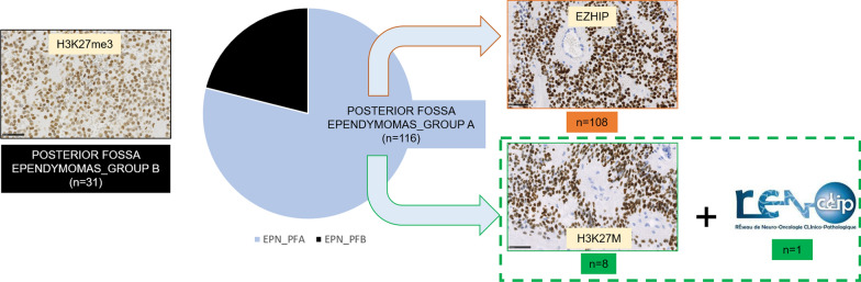
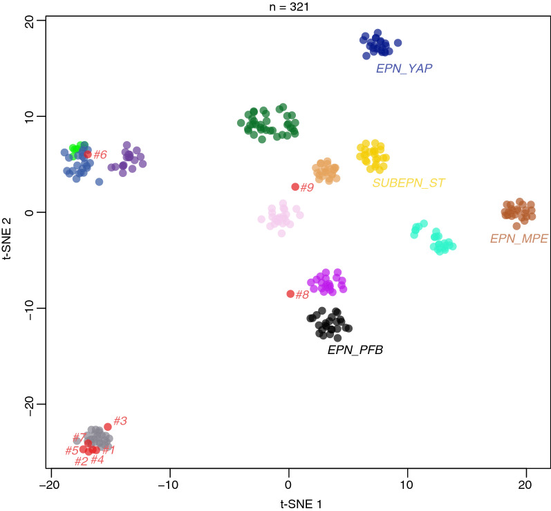
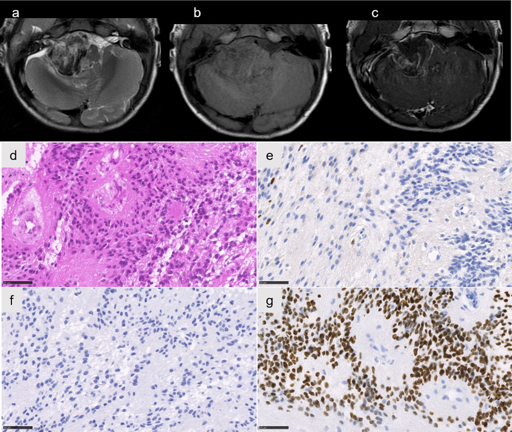
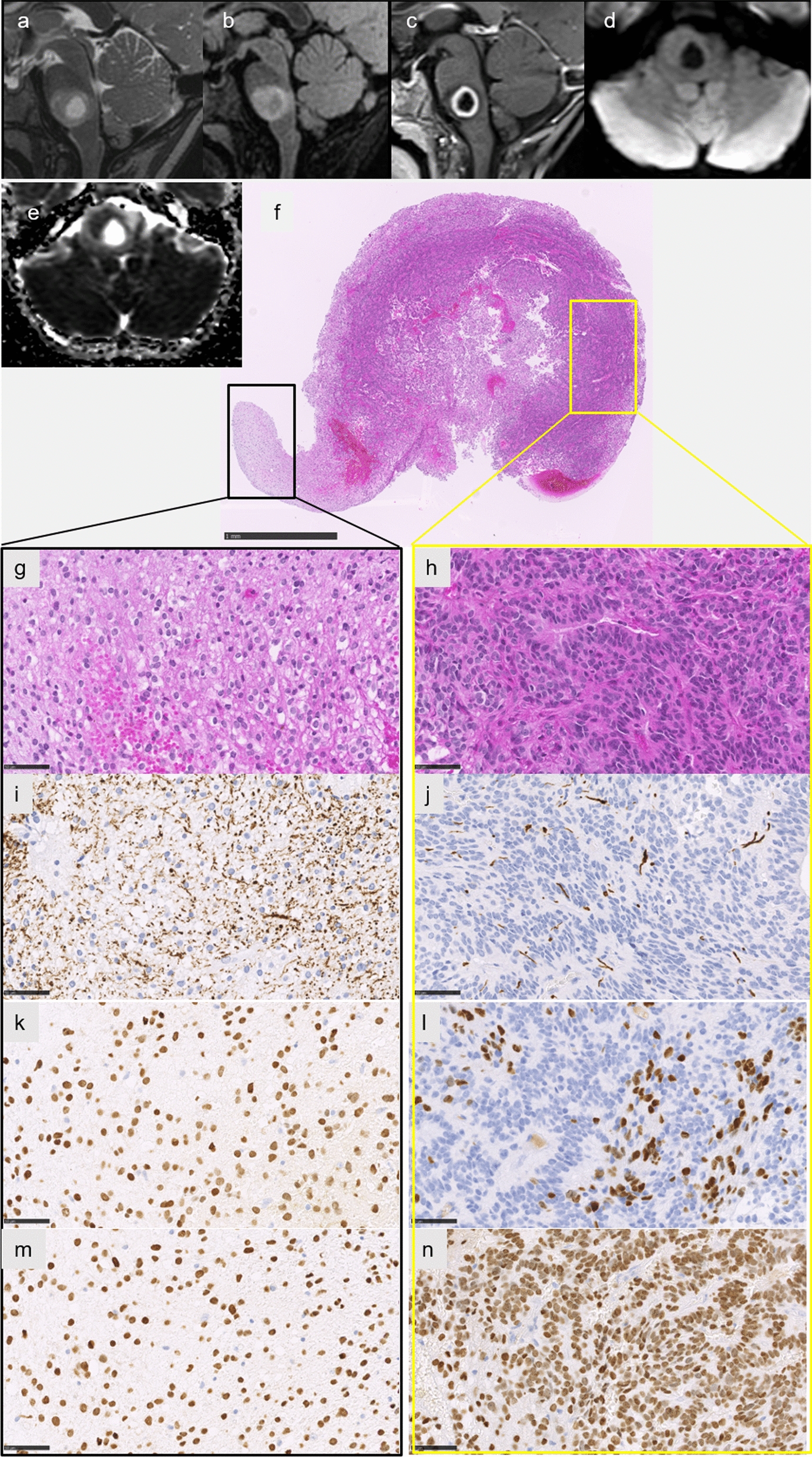
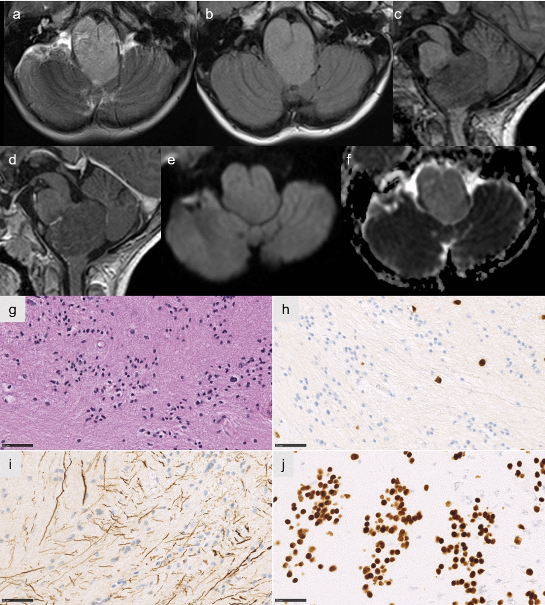
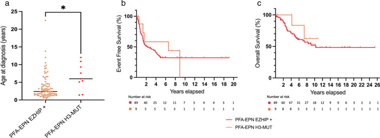
Posterior fossa group A ependymomas (EPN_PFA) are characterized by a loss of H3 K27 trimethylation due to either EZHIP overexpression or H3 p.K27M mutation, similar to H3 K27-altered diffuse midline gliomas (DMG), but in reverse proportions. Very little data is available in the literature concerning H3 K27M-mutant EPN_PFA. Here, we retrospectively studied a series of nine pediatric tumors initially diagnosed as H3 K27M-mutant EPN_PFA to compare them to EZHIP-overexpressing EPN_PFA in terms of radiology, follow-up, histopathology, and molecular biology (including DNA-methylation profiling). Seven tumors clustered within EPN_PFA by DNA-methylation analysis and t-distributed stochastic neighbor embedding. Among the two remaining cases, one was reclassified as a DMG and the last was unclassified. H3 K27M-mutant EPN_PFA cases were significantly older than their counterparts with an EZHIP overexpression. Radiological and histopathological central review of our seven H3 K27M-mutant EPN_PFA cases found them to be similar to their counterparts with an EZHIP overexpression. Sequencing analyses revealed HIST1H3B (n = 2), HIST1H3C (n = 2), H3F3A (n = 1), and HIST1H3D (n = 1) K27M mutations (no sequencing analysis available for the last case which was immunopositive for H3K27M). Consequently, HIST1H3C/D mutations are more frequently observed in EPN_PFA than in classic pontine DMG, H3K27-mutant. Overall survival and event-free survival of EZHIP-overexpressing and H3 K27M-mutant EPN_PFA were similar. After surgery and radiation therapy, 5/7 patients were alive at the end of the follow-up. In summary, the diagnosis of EPN_PFA must include tumor location, growth pattern, Olig2 expression, and DNA-methylation profiling before it can be differentiated from DMG, H3 K27-altered.
Keywords: DNA-methylation; Ependymoma; Histones; Posterior fossa.
© 2022. The Author(s).
Conflict of interest statement
The authors declare that they have no conflict of interest.
Figures
References
-
- Alturkustani M, Cotter JA, Mahabir R, Hawes D, Koschmann C, Venneti S, Judkins AR, Szymanski LJ. Autopsy findings of previously described case of diffuse intrinsic pontine glioma-like tumor with EZHIP expression and molecular features of PFA ependymoma. Acta Neuropathol Commun. 2021;9:113. doi: 10.1186/s40478-021-01215-5. - DOI - PMC - PubMed
-
- Bugiani M, van Zanten SEMV, Caretti V, Schellen P, Aronica E, Noske DP, Vandertop WP, Kaspers GJL, van Vuurden DG, Wesseling P, Hulleman E. Deceptive morphologic and epigenetic heterogeneity in diffuse intrinsic pontine glioma. Oncotarget. 2017;8:60447–60452. doi: 10.18632/oncotarget.19726. - DOI - PMC - PubMed
-
- Capper D, Jones DTW, Sill M, Hovestadt V, Schrimpf D, Sturm D, Koelsche C, Sahm F, Chavez L, Reuss DE, Kratz A, Wefers AK, Huang K, Pajtler KW, Schweizer L, Stichel D, Olar A, Engel NW, Lindenberg K, Harter PN, Braczynski A, Plate KH, Dohmen H, Garvalov BK, Coras R, Hölsken A, Hewer E, Bewerunge-Hudler M, Schick M, Fischer R, Beschorner R, Schittenhelm J, Staszewski O, Wani K, Varlet P, Pages M, Temming P, Lohmann D, Selt F, Witt H, Milde T, Witt O, Aronica E, Giangaspero F, Rushing E, Scheurlen W, Geisenberger C, Rodriguez FJ, Becker A, Preusser M, Haberler C, Bjerkvig R, Cryan J, Farrell M, Deckert M, Hench J, Frank S, Serrano J, Kannan K, Tsirigos A, Brück W, Hofer S, Brehmer S, Seiz-Rosenhagen M, Hänggi D, Hans V, Rozsnoki S, Hansford JR, Kohlhof P, Kristensen BW, Lechner M, Lopes B, Mawrin C, Ketter R, Kulozik A, Khatib Z, Heppner F, Koch A, Jouvet A, Keohane C, Mühleisen H, Mueller W, Pohl U, Prinz M, Benner A, Zapatka M, Gottardo NG, Driever PH, Kramm CM, Müller HL, Rutkowski S, von Hoff K, Frühwald MC, Gnekow A, Fleischhack G, Tippelt S, Calaminus G, Monoranu C-M, Perry A, Jones C, Jacques TS, Radlwimmer B, Gessi M, Pietsch T, Schramm J, Schackert G, Westphal M, Reifenberger G, Wesseling P, Weller M, Collins VP, Blümcke I, Bendszus M, Debus J, Huang A, Jabado N, Northcott PA, Paulus W, Gajjar A, Robinson G, Taylor MD, Jaunmuktane Z, Ryzhova M, Platten M, Unterberg A, Wick W, Karajannis MA, Mittelbronn M, Acker T, Hartmann C, Aldape K, Schüller U, Buslei R, Lichter P, Kool M, Herold-Mende C, Ellison DW, Hasselblatt M, Snuderl M, Brandner S, Korshunov A, von Deimling A, Pfister SM. DNA methylation-based classification of central nervous system tumours. Nature. 2018;555:469–474. doi: 10.1038/nature26000. - DOI - PMC - PubMed
-
- Castel D, Kergrohen T, Tauziède-Espariat A, Mackay A, Ghermaoui S, Lechapt E, Pfister SM, Kramm CM, Boddaert N, Blauwblomme T, Puget S, Beccaria K, Jones C, Jones DTW, Varlet P, Grill J, Debily M-A. Histone H3 wild-type DIPG/DMG overexpressing EZHIP extend the spectrum diffuse midline gliomas with PRC2 inhibition beyond H3–K27M mutation. Acta Neuropathol (Berl) 2020;139:1109–1113. doi: 10.1007/s00401-020-02142-w. - DOI - PubMed
Publication types
MeSH terms
Substances
Supplementary concepts
LinkOut - more resources
Full Text Sources
Other Literature Sources
Medical
