. 2022 Sep 14;19(1):226.
doi: 10.1186/s12974-022-02587-8.

Captopril alleviates epilepsy and cognitive impairment by attenuation of C3-mediated inflammation and synaptic phagocytosis

Affiliations

Captopril alleviates epilepsy and cognitive impairment by attenuation of C3-mediated inflammation and synaptic phagocytosis

Xinyan Dong et al. J Neuroinflammation. .

Abstract

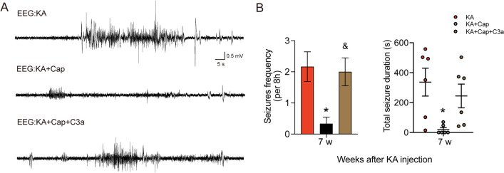
Evidence from experimental and clinical studies implicates immuno-inflammatory responses as playing an important role in epilepsy-induced brain injury. Captopril, an angiotensin-converting enzyme inhibitor (ACEi), has previously been shown to suppress immuno-inflammatory responses in a variety of neurological diseases. However, the therapeutic potential of captopril on epilepsy remains unclear. In the present study, Sprague Dawley (SD) rats were intraperitoneally subjected to kainic acid (KA) to establish a status epilepticus. Captopril (50 mg/kg, i.p.) was administered daily following the KA administration from day 3 to 49. We found that captopril efficiently suppressed the KA-induced epilepsy, as measured by electroencephalography. Moreover, captopril ameliorated the epilepsy-induced cognitive deficits, with improved performance in the Morris water maze, Y-maze and novel objective test. RNA sequencing (RNA-seq) analysis indicated that captopril reversed a wide range of epilepsy-related biological processes, particularly the glial activation, complement system-mediated phagocytosis and the production of inflammatory factors. Interestingly, captopril suppressed the epilepsy-induced activation and abnormal contact between astrocytes and microglia. Immunohistochemical experiments demonstrated that captopril attenuated microglia-dependent synaptic remodeling presumably through C3-C3ar-mediated phagocytosis in the hippocampus. Finally, the above effects of captopril were partially blocked by an intranasal application of recombinant C3a (1.3 μg/kg/day). Our findings demonstrated that captopril reduced the occurrence of epilepsy and cognitive impairment by attenuation of inflammation and C3-mediated synaptic phagocytosis. This approach can easily be adapted to long-term efficacy and safety in clinical practice.

Keywords: Captopril; Cognitive deficits; Complement 3; Epilepsy; Glial activation; Synaptic phagocytosis.


Conflict of interest statement

The authors have no financial conflicts of interest.

Figures

Fig. 1
Fig. 1
Captopril treatment attenuates the KA-induced epilepsy in rats. A The experimental design is shown. B Representative 2 min EEG recording during epilepsy (7 weeks after KA induction) indicates the captopril treatment significantly suppresses the KA-induced recurrent seizures. C, D Quantification of the frequency and total seizure duration validates the captopril treatment significantly suppresses the KA-induced recurrent seizures. n = 6 rats/group, *p < 0.05, **p < 0.01, Student’s t test. Data are presented as mean ± SE
Fig. 2
Fig. 2
Captopril treatment ameliorates epilepsy-related memory impairment in rats. A The experiment diagram of NOR is shown. B, C Representative heat map of traveling track diagram around objects and quantification of discrimination index show captopril treatment significantly ameliorates the epilepsy-related cognitive function impairment. D A schematic diagram of Y-maze test is shown. E Quantification of spontaneous alteration rate in Y-maze test shows captopril treatment reverses the epilepsy-related deficits in spatial memory. F The mean escape latency to the hidden platform in the water maze as a short-term learning function of 4 training days shows no significant difference among the groups. n = 6 rats/group, two-way ANOVA followed by Dunnett’s post hoc test. GH Representative swimming paths and quantification of time spent in the platform quadrant on day 5 (spatial probe test day) show captopril treatment significantly improves the epilepsy-impaired short-term memory. n = 6 rats/group, #p < 0.05, ##p < 0.01, compared to control; *p < 0.05, **p < 0.01, compared to KA, one-way ANOVA followed by Tukey’s post hoc test. Data are presented as mean ± SE
Fig. 3
Fig. 3
Captopril treatment reduces the KA-induced phagocytosis and inflammatory responses in the hippocampus. RNA-seq analysis was performed using the hippocampal tissues 9 weeks after the KA induction. Results were normalized to the control group. P-values were obtained by edgeR using an exact test through the negative binomial distribution. A The volcano plot is shown for gene expression in the hippocampus with significantly increased (red) or decreased (blue) expression (p < 0.05, |log2|> 1) compared between the control and KA group (left) or between the KA and KA + Cap group (right). B Hierarchical clustering was performed for all the significantly altered gene expression. Magenta indicates the number of up-regulated genes and blue indicates the number of down-regulated genes. C The number of the shared and distinct DEGs in the hippocampus in the KA and KA + Cap groups. D GO and KEGG analysis of the transcripts with the shared DEGs in the KA and KA + Cap groups are shown. E The STRING analysis of the interactions of 240 top-regulated genes recovered after captopril treatment is shown. n = 3 rats/group
Fig. 4
Fig. 4
Captopril treatment suppresses the genes expression within the inflammation and phagocytosis. A Gene set enrichment analysis (GSEA) indicates captopril treatment recovers the KA-induced down-regulation of gene expression regarding cytokine–cytokine receptor interaction, phagocytosis, complement and coagulation cascades in the hippocampus. BD The heat map shows the top-regulated gene expression of phagocytosis, complement and coagulation cascades, and cytokine–cytokine receptor interaction in the hippocampus in the KA and KA + Cap groups. E Captopril treatment significantly reduces the gene expression related to inflammatory and immunological regulation in the hippocampus compared to the KA group. n = 6 rats/group, #p < 0.05, ##p < 0.01, ###p < 0.001, compared to control; *p < 0.05, **p < 0.01, ***p < 0.001, compared to KA, one-way ANOVA followed by Tukey’s post hoc test. Data are presented as mean ± SE
Fig. 5
Fig. 5
Captopril treatment suppresses the KA-induced glial cell activation in the hippocampus. A Captopril treatment significantly reduced the KA-induced gene expression of glial markers in the hippocampus 9 weeks after the induction. n = 6 rats/group. B Representative images show Iba1 (cyan) and Gfap (red) staining in the control, KA and KA + Cap groups. Scale bar = 50 μm. C Quantitative analysis shows that captopril treatment significantly attenuates the KA-induced upregulation of the number of Iba1 (cyan) and Gfap (red) positive glial cells in the hippocampal CA1. D Quantitative analysis shows captopril treatment significantly attenuates the KA-induced upregulation of Iba1 (cyan) and Gfap (red) intensity in the hippocampal CA1, n = 3 images from 3 rats/group. E Representative images show the microglial morphology (Iba1, cyan; transformed skeleton, gray) in the control, KA and KA + Cap groups. Scale bar = 5 μm. F Quantification of the branch number (top) and length (bottom) of the microglia process shows that captopril treatment significantly attenuates the epilepsy-induced microglia activation compared to the KA group. n = 12 cells from 3 rats/group, #p < 0.05, ##p < 0.01, ###p < 0.001, compared to control; *p < 0.05, **p < 0.01, ***p < 0.001, compared to KA, one-way ANOVA followed by Tukey’s post hoc test. Data are expressed as mean ± SE
Fig. 6
Fig. 6
Captopril treatment suppresses the KA-induced contact between glial cells in the hippocampus. A Representative anti-Iba1 and anti-Gfap double immunostaining indicates that captopril treatment reduces the KA-induced contact between astrocytes and microglia. Scale bar = 5 μm. B Quantification of Gfap (red) signal intensity around microglia (cyan) is shown. n = 10–12 cells from 3 rats/group, ###p < 0.001, compared to control; ***p < 0.001, compared to KA, one-way ANOVA followed by Tukey’s post hoc test. Data are expressed as mean ± SE
Fig. 7
Fig. 7
Captopril treatment attenuates the KA-induced C3 expression in astrocytes and C3ar expression in microglia in the hippocampus 9 weeks after the KA induction. A Captopril significantly reduces the KA-induced upregulation of mRNA expression of C3 and C3ar in the hippocampus. n = 6 rats/group. B Representative anti-C3 (green) and anti-Gfap (red) double immunostaining indicates captopril attenuates the KA-induced elevation in astrocytic C3 production. C Representative anti-C3 (green) and anti-Iba1 (cyan) double immunostaining indicates C3 is not expressed in microglia in the hippocampus. D Representative anti-C3ar (red) and anti-Iba1 (cyan) double immunostaining indicates captopril attenuates the KA-induced elevation in microglia C3ar expression. E Quantification of C3 (green) signal intensity within Gfap+ (red) astrocytes is shown. F Quantification of C3ar (red) signal intensity within Iba1 (cyan) microglia is shown. Scale bar = 10 μm. n = 9 images from 3 rats/group, ##p < 0.01, ###p < 0.001, compared to control; *p < 0.05, **p < 0.01; ***p < 0.001, compared to KA, one-way ANOVA followed by Tukey’s post hoc test. Data are expressed as mean ± SE
Fig. 8
Fig. 8
Captopril treatment reduces the KA-induced synaptic phagocytosis by activated microglia. A Immunostaining of Cd68 (green), synapsin (red) and Iba1 (cyan) in the hippocampal CA1 indicates captopril significantly attenuates the KA-induced synaptic pruning by microglia. Top, scale bar = 5 μm. Bottom, scale bar = 10 μm. B Quantification of Cd68 and synapsin immunofluorescent signals within Iba1+ microglia indicates captopril treatment significantly reduces the KA-induced microglia engulfment capacity as measured by lysosomal content within each microglia (CD68 immunoreactivity per cell). C Quantification shows that captopril reduces the KA-induced upregulation of the proportion of microglia with Cd68+ (green) signal in the hippocampal CA1. D Representative confocal images of synapsin (red) from pyramidale and radiatum in the hippocampal CA1 are shown. Scale bar = 5 μm. E Quantification of punctuated integrated signal intensity of synapsin indicates captopril significantly attenuates the KA-induced synaptic loss in the pyramidale and radiatum layer of hippocampal CA1. Scale bar = 5 μm. n = 9 images from 3 rats/group, ###p < 0.001, compared to control; ***p < 0.001, compared to KA, one-way ANOVA followed by Tukey’s post hoc test. Data are presented as mean ± SE
Fig. 9
Fig. 9
Nasal instillation of C3a aggravates epilepsy-related cognitive dysfunction after captopril treatment. A The experimental design is shown. B Representative heat maps of the duration the rat spent around the old and new objects are shown. C C3a nasal drip reverses the cognitive amelioration by captopril treatment in NOR test. D A schematic diagram of Y-maze test is shown. E C3a nasal drip reverses the spatial memory amelioration from captopril in spontaneous alternation in Y-maze task. F The mean escape latency to the hidden platform in the water maze as a short-term learning function of 4 training days shows no significant difference. n = 6 rats/group, two-way ANOVA followed by Dunnett’s post hoc test. G Representative swimming paths in the MWM test for day 5 (spatial probe test day) are shown. H C3a nasal drip significantly reverses the therapeutic effects of captopril on the short-term memory impairment, indicated by a reduction in the percentage of time spent in the target quadrant where the platform is located. n = 6 rats/group, #p < 0.05, ##p < 0.01, compared to control; *p < 0.05, **p < 0.01, compared to KA + Cap; &p < 0.05; &&p < 0.01, compared to KA + Cap, one-way ANOVA followed by Tukey’s post hoc test. Data are presented as mean ± SE
Fig. 10
Fig. 10
Intranasal instillation of C3a promotes epileptogenesis after captopril treatment in the KA-induced model of epilepsy. A Representative 2-min EEG recording during epilepsy (7 weeks after KA induction) from the KA, KA + Cap, and KA + Cap + C3a groups are shown. B C3a nasal drip significantly elevates the frequency and total duration of seizures compared to the KA + Cap group 7 weeks after the KA induction. n = 6 rats/group, *p < 0.05, compared to KA; &p < 0.05, compared to KA + Cap, one-way ANOVA followed by Tukey’s post hoc test. Data are presented as mean ± SE
Fig. 11
Fig. 11
Intranasal instillation of C3a promotes the microglia-mediated synaptic phagocytosis after captopril treatment. A Representative anti-C3ar (red) and anti-Iba1 (cyan) double immunostaining indicates that C3a nasal drip reverses the C3ar suppression in microglia following captopril treatment. Scale bar = 10 μm. B Quantification of C3ar (red) signal intensity within Iba1+ (cyan) microglia is shown. C, D Immunostaining of Cd68 (green), synapsin (red) and Iba1 (cyan) and quantification of microglia with Cd68+ signal in the hippocampal CA1 indicate that C3a significantly reverses the inhibitory effects of captopril on synaptic pruning by microglia. Scale bar = 10 μm. E Representative confocal images of synapsin (red) from pyramidale and radiatum layer in the hippocampal CA1 are shown. Scale bar = 5 μm. F Quantification of integrated signal intensity synapsin indicates C3a significantly reverses the therapeutic effects of captopril treatment on reducing synaptic loss in the pyramidale and radiatum layer in the hippocampal CA1. n = 9 images from 3 rats/group, ***p < 0.001, compared to KA; &p < 0.05, &&&p < 0.001, compared to KA + Cap, one-way ANOVA followed by Tukey’s post hoc test. Data are presented as mean ± SE

Comment in

References

    1. Devinsky O, Vezzani A, O'Brien TJ, Jette N, Scheffer IE, de Curtis M, Perucca P. Epilepsy. Nat Rev Dis Primers. 2018;4:18024. doi: 10.1038/nrdp.2018.24. - DOI - PubMed
    1. Tellez-Zenteno JF, Hernandez-Ronquillo L. A review of the epidemiology of temporal lobe epilepsy. Epilepsy Res Treat. 2012;2012:630853. - PMC - PubMed
    1. Sen A, Capelli V, Husain M. Cognition and dementia in older patients with epilepsy. Brain. 2018;141:1592–1608. doi: 10.1093/brain/awy022. - DOI - PMC - PubMed
    1. Perucca E, Brodie MJ, Kwan P, Tomson T. 30 years of second-generation antiseizure medications: impact and future perspectives. Lancet Neurol. 2020 doi: 10.1016/S1474-4422(20)30035-1. - DOI - PubMed
    1. Loscher W, Klitgaard H, Twyman RE, Schmidt D. New avenues for anti-epileptic drug discovery and development. Nat Rev Drug Discov. 2013;12:757–776. doi: 10.1038/nrd4126. - DOI - PubMed