

Insight of vitellogenesis patterns: A comparative analysis of the differences between the primary and secondary vitellogenesis period in the ovary, hepatopancreas, and muscle of mud crab, scylla paramamosain
- PMID: 36105103
- PMCID: PMC9465302
- DOI: 10.3389/fgene.2022.965070
Insight of vitellogenesis patterns: A comparative analysis of the differences between the primary and secondary vitellogenesis period in the ovary, hepatopancreas, and muscle of mud crab, scylla paramamosain
Abstract
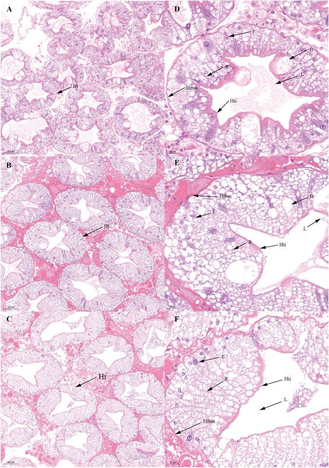
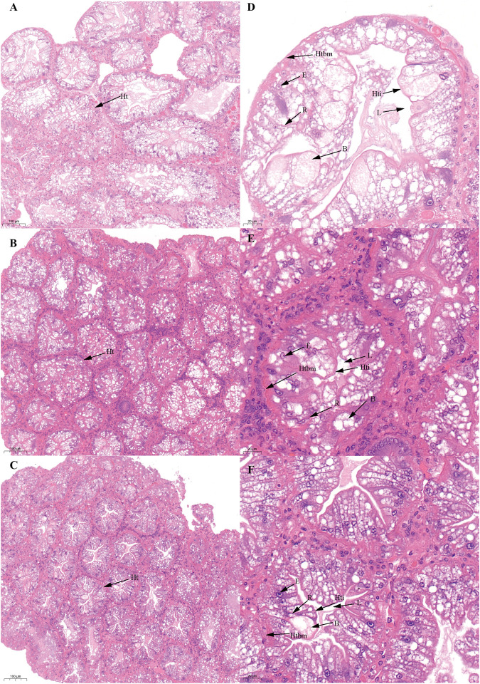
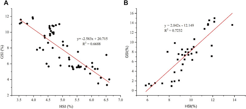
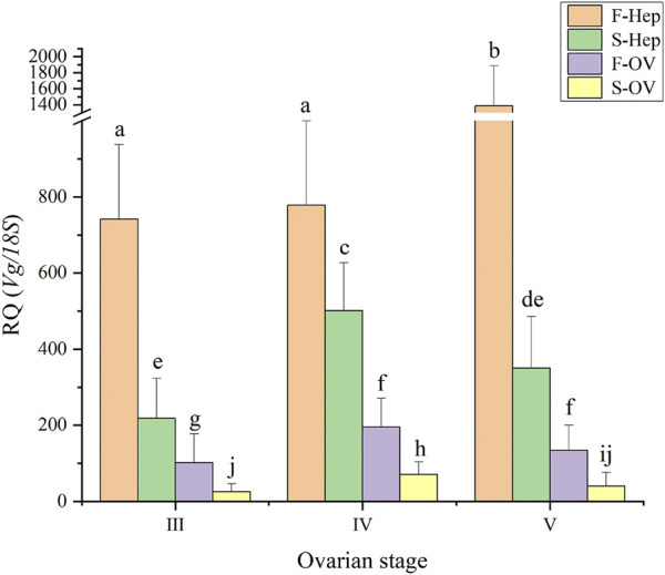
The mud crab, Scylla paramamosain, has abundant nutrients in its edible parts, ovary, hepatopancreas, and muscle during the ovarian maturation stage. The ovary of S. paramamosain can re-mature after spawning during the secondary ovarian maturation period. We aimed to analyze the characteristics of the first vitellogenesis period (FVP) and second vitellogenesis period (SVP) of S. paramamosain during ovarian maturation to understand the differences in vitellogenesis patterns between the first and second ovarian maturation periods. Accordingly, the gonadosomatic index (GSI) and hepatopancreatic index (HSI), the external and histological characteristics of the ovary and hepatopancreas, the Sp-Vg (vitellogenin, Vg) expression levels in the hepatopancreas and ovary, and the dynamics of the biochemical components in the ovary, hepatopancreas, and muscle were determined. Based on the results, the GSI was significantly positively correlated with HSI during the FVP and significantly negatively correlated with HSI from stage Ⅳ to stage Ⅴ of the SVP. A significant difference was found between the FVP and SVP in the hepatopancreas. Notably, the hepatopancreas displayed a gradual degeneration trend during the SVP. The expression level of Sp-Vg was significantly higher in the hepatopancreas than that in the ovary during the FVP and SVP. Seventeen amino acids were detected in the hepatopancreas, ovary, and muscle during the FVP and SVP, with glutamate as the predominant amino acid. During the FVP and SVP, the C16:0 and C18:1n9c were the dominant fatty acids in the hepatopancreas and ovary, the MUFA gradually increased in the ovary and hepatopancreas, and a significant difference was found in the dynamic trend of the HUFA and SFA contents from stage Ⅳ to stage Ⅴ between the FVP and SVP. These findings indicate that the ovary can re-mature after spawning in S. paramamosain and can maintain the status of the first ovarian maturation; however, the hepatopancreas gradually degenerate during the SVP.
Keywords: GSI and HSI; Scylla paramamosain; biochemical components; ovarian development; ovarian re-maturation; vitellogenesis patterns.
Copyright © 2022 Ren, Wang, Liu, Luo, Fu, Zhang, Ma, Zhao, Chen, Jiang and Ma.
Conflict of interest statement
The authors declare that the research was conducted in the absence of any commercial or financial relationships that could be construed as a potential conflict of interest.
Figures


Similar articles
-
Role of neuroparsin 1 in vitellogenesis in the mud crab, Scylla paramamosain.Gen Comp Endocrinol. 2020 Jan 1;285:113248. doi: 10.1016/j.ygcen.2019.113248. Epub 2019 Aug 17. Gen Comp Endocrinol. 2020. PMID: 31430448
-
Tissue-specific vitellogenesis and 17β-estradiol facilitate ovarian maturation of the swimming crab Portunus trituberculatus.Comp Biochem Physiol A Mol Integr Physiol. 2025 Mar;301:111798. doi: 10.1016/j.cbpa.2024.111798. Epub 2024 Dec 31. Comp Biochem Physiol A Mol Integr Physiol. 2025. PMID: 39746648
-
The mechanism of regulation of ovarian maturation by red pigment concentrating hormone in the mud crab Scylla paramamosain.Anim Reprod Sci. 2016 Jan;164:152-61. doi: 10.1016/j.anireprosci.2015.11.025. Epub 2015 Dec 3. Anim Reprod Sci. 2016. PMID: 26679434
-
Morphological, biochemical and histological analysis of mud crab ovary and hepatopancreas at different stages of development.Anim Reprod Sci. 2018 Aug;195:274-283. doi: 10.1016/j.anireprosci.2018.06.005. Epub 2018 Jun 9. Anim Reprod Sci. 2018. PMID: 29910008
-
Evaluation of the Effect of Adipokinetic Hormone/Corazonin-Related Peptide (ACP) on Ovarian Development in the Mud Crab, Scylla paramamosain.Animals (Basel). 2024 Dec 23;14(24):3706. doi: 10.3390/ani14243706. Animals (Basel). 2024. PMID: 39765610 Free PMC article.
Cited by
-
Functional complementation of two splicing variants of Gustavus in Neocaridina denticulata sinensis during ovarian maturation.Sci Rep. 2024 Sep 9;14(1):20939. doi: 10.1038/s41598-024-72080-0. Sci Rep. 2024. PMID: 39251721 Free PMC article.
-
Temporal and Spatial Signatures of Scylla paramamosain Transcriptome Reveal Mechanistic Insights into Endogenous Ovarian Maturation under Risk of Starvation.Int J Mol Sci. 2024 Jan 5;25(2):700. doi: 10.3390/ijms25020700. Int J Mol Sci. 2024. PMID: 38255774 Free PMC article.
-
Reproductive Dynamics of Spot Tail Mantis Shrimp (Squilla mantis): Insights from the Central Mediterranean Sea.Animals (Basel). 2024 Aug 28;14(17):2503. doi: 10.3390/ani14172503. Animals (Basel). 2024. PMID: 39272288 Free PMC article.
-
Morphometrics and Reproductive Characteristics of the Freshwater Crab Sartoriana spinigera from the Habitat of Ratargul Swamp Forest, Bangladesh: An Approach to Conservation.Scientifica (Cairo). 2024 Aug 21;2024:4550875. doi: 10.1155/2024/4550875. eCollection 2024. Scientifica (Cairo). 2024. PMID: 39206113 Free PMC article.
References
-
- Alava V. R., Quinitio E. T., De Pedro J. B., Orosco Z. G., Wille M. (2007). Reproductive performance, lipids and fatty acids of mud crab Scylla serrata (Forsskål) fed dietary lipid levels. Aquac. Res. 38 (14), 1442–1451. 10.1111/j.1365-2109.2007.01722.x - DOI
-
- Bureau of Fisheries Ministry of Agriculture PRC (2021). China Fishery statistical Yearbook. Beijing, China: Chinese Agriculture Press.
LinkOut - more resources
Full Text Sources