

Analysis of Causes and Results of Fetal Growth in Utero Caused by Genetic Factors Detected by Ultrasound
- PMID: 36105440
- PMCID: PMC9452974
- DOI: 10.1155/2022/3703132
Analysis of Causes and Results of Fetal Growth in Utero Caused by Genetic Factors Detected by Ultrasound
Retraction in
-
Retracted: Analysis of Causes and Results of Fetal Growth in Utero Caused by Genetic Factors Detected by Ultrasound.Contrast Media Mol Imaging. 2023 Jul 12;2023:9816748. doi: 10.1155/2023/9816748. eCollection 2023. Contrast Media Mol Imaging. 2023. PMID: 37476455 Free PMC article.
Abstract
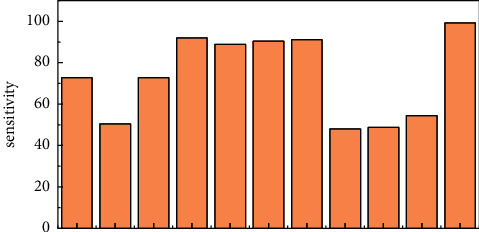
In order to investigate the value of the ultrasonic monitoring of maternal and fetal vascular parameters, serum vitamin D, and placental growth factor (PLGF) in predicting fetal growth restriction (FGR), a method of ultrasonic detection of genetic factors causing fetal growth in utero was proposed. 125 pregnant women with FGR diagnosed in our hospital from June 2018 to June 2021 (the FGR group) and 125 pregnant women with a normal prenatal examination (the control group) were collected retrospectively. The systolic/diastolic blood flow ratio (S/D), pulsatile index (PI), and resistance index (RI) of the fetal umbilical artery (UA), middle cerebral artery (MCA), and maternal uterine artery (UtA) were monitored by ultrasound at 20 to 24 weeks of gestation, and the levels of serum vitamin D and PLGF were detected. The receiver operating characteristic curve (ROC curve) was used to evaluate the predictive value of FGR. The results showed that the S/D, PI, and RI of UA in the FGR group were higher than those in the control group, the areas under the curve (AUC) were 0.866, 0.817, and 0.849, and the sensitivity and specificity were (72.8%, 91.2%), (50.4%, 100%), and (72.8%, 91.2%), respectively. The S/D, PI, and RI of MCA were lower than those of the control group. The AUC was 0.882, 0.869, and 0.834, respectively; the sensitivity and specificity were (92.0%, 74.4%), (88.8%, 81.6%), and (90.4%, 72%), respectively. The S/D, PI, and RI of UtA were higher than those of the control group; the AUC was 0.768, 0.729, and 0.732; the sensitivity and specificity were (91.2%, 52%), (48%, 90.4%), and (48.8%, 90.4%), respectively. The serum levels of vitamin D and PLGF were lower than those of the control group (AUC 0.784 and 0.807), and the sensitivity and specificity were (54.4%, 91.2%) and (99.2%, 52%), respectively. It was concluded that the ultrasound monitoring of UA, MCA, and UtA in pregnant women in the middle of pregnancy and detection of serum vitamin D and PLGF levels had a certain predictive value for FGR. Moreover, the comprehensive evaluation could reduce the occurrence of FGR in high-risk pregnant women.
Copyright © 2022 Mei Yu et al.
Conflict of interest statement
The authors declare that they have no conflicts of interest.
Figures
Similar articles
-
Role of Doppler ultrasound at time of diagnosis of late-onset fetal growth restriction in predicting adverse perinatal outcome: prospective cohort study.Ultrasound Obstet Gynecol. 2020 Jun;55(6):793-798. doi: 10.1002/uog.20406. Epub 2020 May 8. Ultrasound Obstet Gynecol. 2020. PMID: 31343783
-
Fetal and Neonatal Middle Cerebral Artery Hemodynamic Changes and Significance under Ultrasound Detection in Hypertensive Disorder Complicating Pregnancy Patients with Different Severities.Comput Math Methods Med. 2022 Jun 28;2022:6110228. doi: 10.1155/2022/6110228. eCollection 2022. Comput Math Methods Med. 2022. PMID: 35799667 Free PMC article.
-
Prenatal Prediction of Fetal Growth Restriction and Postnatal Outcomes by Ultrasound Assessment of Fetal Myocardial Performance Index and Blood Flow Spectrum.Evid Based Complement Alternat Med. 2022 May 5;2022:4234137. doi: 10.1155/2022/4234137. eCollection 2022. Evid Based Complement Alternat Med. 2022. Retraction in: Evid Based Complement Alternat Med. 2023 Jun 21;2023:9841616. doi: 10.1155/2023/9841616. PMID: 35571730 Free PMC article. Retracted.
-
Cerebral-placental-uterine ratio as novel predictor of late fetal growth restriction: prospective cohort study.Ultrasound Obstet Gynecol. 2019 Sep;54(3):367-375. doi: 10.1002/uog.20150. Ultrasound Obstet Gynecol. 2019. PMID: 30338593
-
Correlation between Parturients' Uterine Artery Blood Flow Spectra in the First and Second Trimesters of Pregnancy and Fetal Growth Restriction.J Healthc Eng. 2021 Dec 14;2021:2129201. doi: 10.1155/2021/2129201. eCollection 2021. J Healthc Eng. 2021. PMID: 34950439 Free PMC article.
Cited by
-
Retracted: Analysis of Causes and Results of Fetal Growth in Utero Caused by Genetic Factors Detected by Ultrasound.Contrast Media Mol Imaging. 2023 Jul 12;2023:9816748. doi: 10.1155/2023/9816748. eCollection 2023. Contrast Media Mol Imaging. 2023. PMID: 37476455 Free PMC article.
References
-
- Meng D. H., Zhang Y., Ma S. S., et al. The role of parathyroid hormone during pregnancy on the relationship between maternal vitamin d deficiency and fetal growth restriction: a prospective birth cohort study. British Journal of Nutrition . 2020;124(4):432–439. doi: 10.1017/s0007114520001105. - DOI - PubMed
Publication types
MeSH terms
Substances
LinkOut - more resources
Full Text Sources
Research Materials
Miscellaneous
