. 2022 Aug 15;12(35):22826-22842.
doi: 10.1039/d2ra02849b. eCollection 2022 Aug 10.

Mutations responsible for the carbapenemase activity of SME-1

Affiliations

Mutations responsible for the carbapenemase activity of SME-1

Vidhu Agarwal et al. RSC Adv. .

Abstract

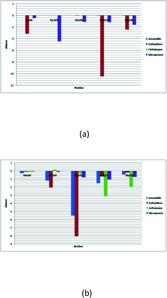
SME-1 is a carbapenemase, produced by Serratia marcescens organism and causes nosocomial infections such as in bloodstream, wounds, urinary tract, or respiratory tract infections. Treatment of such infections becomes very complex due its resistance towards penicillins, cephalosporins, monobactams, and carbapenems. Resistance to such antibiotics is of great medical concern. The misuse and overuse of these antibiotics result in the clinical mutation and production of novel β-lactamase enzymes such as SME-1, which show resistance to carbapenems. Class A contains most of the clinically significant extended spectrum of β-lactamase enzymes and carbapenemases. In this study, class A β-lactamase SME-1 sequence, structure, and binding were compared with naturally mutated class A β-lactamase enzymes and a wild-type TEM-1. This study was performed for revealing mutations, which could be responsible for the carbapenemase activity of SME-1. The dynamic characteristics of SME-1 enzymes manifest a different degree of conservation and variability, which confers them to possess carbapenemase activities. Met69Cys, Glu104Tyr, Tyr105His, Ala237Ser, and Gly238Cys mutations occur in SME-1 as compared to wild-type TEM-1. These mutated residues are present close to active site residues such as Ser70, Lys73, Ser130, Asn132, Glu166, and Asn170, which participate in the hydrolytic reaction of β-lactam antibiotics. Furthermore, these mutated residues demonstrate altered interactions with the β-lactam antibiotics (results in altered binding) and within themselves (results in active site structure alterations), which results in expanding the spectrum of activity of these enzymes. This study provides important insights into the structure and activity relationship of SME-1 enzymes. This is evident from the Ω-loop structure modification, which forms the wall of the active site and repositioning of residues involved in hydrolytic reactions, when present in the complex with meropenem in a stable state of MD simulation at 50 ns. Hence, Met69Cys, Glu104Tyr, Tyr105His, Ala237Ser, and Gly238Cys mutations could result in an altered active site structure, binding, and activity of SME-1 with meropenem and thus become resistantant against meropenem, which is a carbapenem.


Conflict of interest statement

There are no conflicts to declare.

Figures

Fig. 1
Fig. 1. (a) Multiple sequence alignments of SME-1 (PDB ID: 1DY6) along with 13 naturally mutated class A β-lactamase enzymes (TEM-52 (PDB ID: 1HTZ), SHV-1 (PDB ID: 1SHV), SME-1 (PDB ID: 1DY6), NMC-A (PDB ID: 1BUE), SED-1 (PDB ID: 3BFE), CTXM-9 (PDB ID: 1YLJ), CTXM-27 (PDB ID: 1YLP), PENI (PDB ID: 3W4P), GES-2 (PDB ID: 3NI9), GES-11 (PDB ID: 3V3R), GES5 (PDB ID: 4GNU), PER-1 (PDB ID: 1E25) and PER-2 (PDB ID: 4D2O)) and TEM-1 (PDB ID: 1ZG4). (b) Shows the phylogenetic tree for the (PDB ID: 1DY6) along with 13 naturally mutated class A β-lactamase enzymes and TEM-1 (PDB ID: 1ZG4). (c) Shows overall and Ω-loop structure comparison of 14 naturally mutated class A β-lactamase enzymes (including SME-1) with wild type class A β-lactamase enzyme TEM-1. (d) Shows the comparison of SME-1 (carbapenemase), SHV-1 (penicillinase) and TEM-1 (wild type) class A β-lactamase enzyme.
Fig. 2
Fig. 2. Shows the interaction diagram after docking between (a) SME-1_Amoxicillin (b) SME-1_Ceftazidime (c) SME-1_Ceftolozane (d) SME-1_Meropenem (e) SHV-1_Amoxicillin (f) SHV-1_Ceftazidime (g) SHV-1_Ceftolozane (h) SHV-1_Meropenem (i) TEM-1_Amoxicillin (j) TEM-1_Ceftazidime (k) TEM-1_Ceftolozane (l) TEM-1_Meropenem.
Fig. 3
Fig. 3. The RMSD plot of clinically mutated class A β-lactamase enzymes (SME-1 and SHV-1) and wild class A β-lactamase enzyme TEM-1 in complex with (a) amoxicillin (b) ceftazidime (c) ceftolozane (d) meropenem.
Fig. 4
Fig. 4. Shows the RMSF plot and B factor plot of (a) SME-1_Amoxicillin (b) SME-1_Ceftazidime (c) SME-1_Ceftolozane (d) SME-1_Meropenem (e) SHV-1_Amoxicillin (f) SHV-1_Ceftazidime (g) SHV-1_Ceftolozane (h) SHV-1_Meropenem (i) TEM-1_Amoxicillin (j) TEM-1_Ceftazidime (k) TEM-1_Ceftolozane (l) TEM-1_Meropenem complex. Alpha helix (represented by the orange background of the plot), and beta-sheet (represented by the light blue background of the plot) are the loop regions (represented by the white background of the plot). Green lines show the residues of protein that interact with the ligand.
Fig. 5
Fig. 5. The interaction fraction plot of amino acids between (a) SME-1_Amoxicillin (b) SME-1_Ceftazidime (c) SME-1_Ceftolozane (d) SME-1_Meropenem (e) SHV-1_Amoxicillin (f) SHV-1_Ceftazidime (g) SHV-1_Ceftolozane (h) SHV-1_Meropenem (i) TEM-1_Amoxicillin (j) TEM-1_Ceftazidime (k) TEM-1_Ceftolozane (l) TEM-1_Meropenem.
Fig. 6
Fig. 6. Shows the timeline representation of interaction and contact between (a) SME-1_Amoxicillin (b) SME-1_Ceftazidime (c) SME-1_Ceftolozane (d) SME-1_Meropenem (e) SHV-1_Amoxicillin (f) SHV-1_Ceftazidime (g) SHV-1_Ceftolozane (h) SHV-1_Meropenem (i) TEM-1_Amoxicillin (j) TEM-1_Ceftazidime (k) TEM-1_Ceftolozane (l) TEM-1_Meropenem.
Fig. 7
Fig. 7. The principal component analysis of clinically mutated class A β-lactamase enzymes (SME-1 and SHV-1) and wild class A β-lactamase enzyme TEM-1 with (a) amoxicillin (b) ceftazidime (c) ceftolozane (d) meropenem.
Fig. 8
Fig. 8. (a) Shows the residue interaction network of the SME-1 enzyme (b) Shows the residue interaction network of TEM-1 enzyme. The green colour shows mutated residues along with the residues of catalytic importance and the blue colour shows its neighbouring residues that directly interact with mutated residues.
Fig. 9
Fig. 9. (a) Shows the residue decomposition analysis for (a) clinically mutated class A β-lactamase enzyme SME-1 and (b) wild class A β-lactamase enzyme TEM-1.
Fig. 10
Fig. 10. (a) Shows residues that are involved in the hydrolytic reaction with β-lactam antibiotics and are conserved (black), mutated in SME-1 (cyan), SHV-1 (green) and TEM-1 (magenta), at 50 ns (stable state of MD simulation) (b) Shows modifications in Ω-loop structure at 50 ns (stable state of MD simulation).

References

    1. Hammoudi Halat D. Ayoub Moubareck C. Antibiotics. 2020;9:186. doi: 10.3390/antibiotics9040186. - DOI - PMC - PubMed
    1. Biagi M. Shajee A. Vialichka A. Jurkovic M. Tan X. Wenzler E. Antimicrob. Agents Chemother. 2020;64:4. - PMC - PubMed
    1. Khanna A. JCDR. 2013;7:2. - PMC - PubMed
    1. Teklu D. S. Negeri A. A. Legese M. H. Bedada T. L. Woldemariam H. K. Tullu K. D. Antimicrobial Resistance and Infection Control. 2019;8:39. doi: 10.1186/s13756-019-0488-4. - DOI - PMC - PubMed
    1. Sougakoff W. L'Hermite G. Pernot L. Naas T. Guillet V. Nordmann P. Jarlier V. Delettré J. Acta Crystallogr., Sect. D: Biol. Crystallogr. 2002;58:267–274. doi: 10.1107/S0907444901019606. - DOI - PubMed