. 2022 Aug 29:16:981426.
doi: 10.3389/fnana.2022.981426. eCollection 2022.

Innervation of human superficial fascia

Affiliations

Innervation of human superficial fascia

Caterina Fede et al. Front Neuroanat. .

Abstract

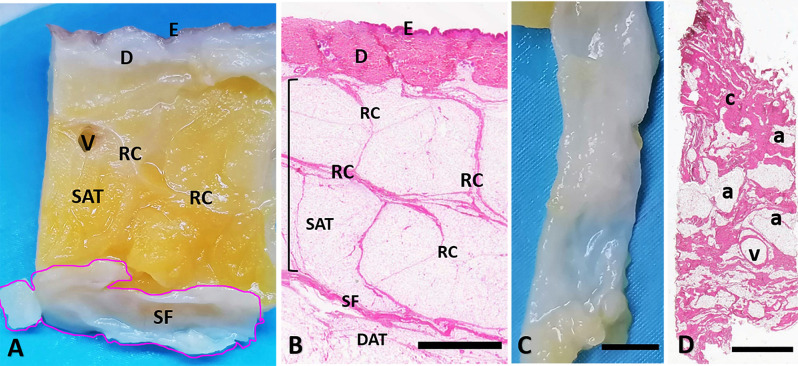
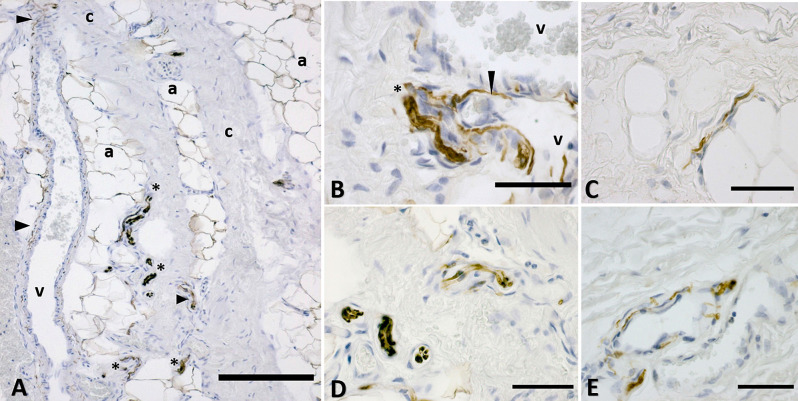
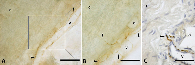
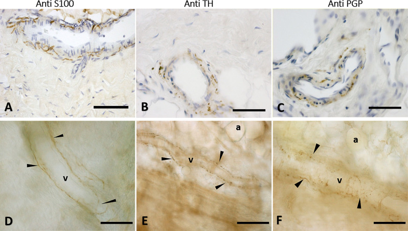
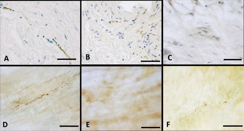
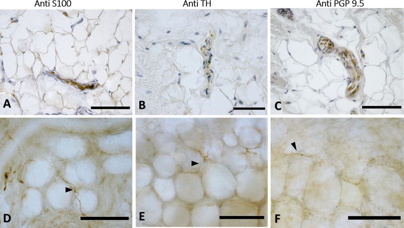
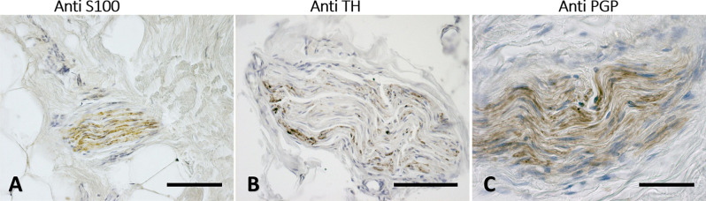
The superficial fascia has only recently been recognized as a specific anatomical structure. Furthermore, whereas it is actually recognized that the innervation of the deep/muscular fascia plays a key role in proprioception and nociception, there are very few studies that have analyzed these characteristics in the superficial fascia. In this work, our group analyzed two different anatomical districts (abdomen and thigh), from volunteer patients, undergoing surgery procedures. Each sample was processed for histological analysis by Hematoxylin&Eosin, and by immunohistochemistry stainings (in 5-micron-paraffin embedded section and in cryosectioned free floating samples), with antibodies specific for nerve fibers: S100 antibody for myelinating and non-myelinating Schwann cells, PGP9.5 antibody as pan-neuronal marker, tyrosine hydroxylase for autonomic innervation. The results revealed a huge innervation: the nervous structures were found above all around blood vessels and close to adipocytes, but they penetrated also in the connective tissue itself and are found in the midst of fibro-adipose tissue. The tissue is pervaded by both thin (mean diameter of 4.8 ± 2.6 μm) and large nerve fiber bundles of greater diameter (21.1 ± 12.2 μm). The ratio S100/TH positivity was equal to 2.96, with a relative percentage of autonomic innervation with of 33.82%. In the light of these findings is evident that the superficial fasciae have a clear and distinct anatomical identity and a specific innervation, which should be considered to better understand their role in thermoregulation, exteroception and pain perception. The knowledge of the superficial fascia may improve grading and developing of different manual approach for treatments of fascial dysfunctions, and the understanding of how some factors like temperature or manual therapies can have an impact on sensitivity of the fascia.

Keywords: autonomic innervation; hypodermis; innervation; nerve fibers; superficial fascia.


Conflict of interest statement

The authors declare that the research was conducted in the absence of any commercial or financial relationships that could be construed as a potential conflict of interest.

Figures

Figure 1
Figure 1
Superficial fascia layer of the abdomen. (A) Formalin-fixed sample of the abdominal region, from skin (E: epidermis, D: dermis) to the superficial fascia (SF). (B) Hematoxylin and eosin stain of all the layers from skin (E: epidermis, D: dermis), to the deep adipose tissue (DAT). The superficial fascia (SF) is the fibrous layer localized between the superficial adipose tissue (SAT, organized in lobules separated by the fibrous septa of the retinacula cutis, RC) and the deep adipose tissue (DAT). The SF highlighted in (A) is isolated from the SAT and the DAT (C) and stained by Hematoxylin and Eosin (D). Panel (D) is a tangential section of the flat embedded SF as in (C). Histologically the SF layer if formed by a net of collagen fibers arranged irregularly (c: connective tissue), interconnected and mixed with adipocytes (a) and crossed by blood vessels (v). Scale Bars = 3 mm.
Figure 2
Figure 2
Distribution of the innervation in superficial fascia: immunohistochemistry by S100 antibody. (A) The connective tissue (c) is crossed by blood vessels (v) and interconnected with areas rich in adipocytes (a). Nervous structures (*indicates big nerves, arrowheads indicate thin nerve fibers) are evident between the collagen fibers, near the adipocytes and in the wall of blood vessels. (B) A nerve (*) and thin nerve fibers (arrow) close to two blood vessels (v). (C) Innervation between the adipocytes area. (D) Nerves crossing the fibrous connective tissue of the SF. (E) Innervation of the wall of a blood vessel supplying the SF. a, adipocyte; v, vessel; c, connective tissue; *, nerve. Arrowheads indicate small nerve fibers. Scale Bars: (A) = 200 μm, (B–E) = 50 μm.
Figure 3
Figure 3
Nerve fibers penetrating the connective tissue. Innervation in a free floating superficial fascia of the abdomen region, by anti-S100 antibody. (A) The wall of the blood vessel (v) is richly innervated, and some nerve fibers enter inside the connective tissue (c) of the SF, as shown in the box, enlarged in (B). Panel (C) shows the same anti-S100 reaction in paraffin-embedded 5 μm section. The arrows indicate the nerve fibers. v, blood vessel; c, connective tissue; a, adipocyte. Scale bars: (A) = 200 μm; (B) = 100 μm; (C) = 50 μm.
Figure 4
Figure 4
Innervation of the blood vessels of superficial fascia. Superficial fascia of the abdomen, stained with S100 (A,D), Tyrosine Hydroxylase (B,E) and PGP9.5 (C,F) antibodies. Panels (A,B,C) are paraffin-embedded-5 μm samples, whereas (D,E,F) are free-floating samples. All the pictures show the innervation of blood vessels. In (E,F) the varicose nature of the axons is evident. v, blood vessel; a, adipocyte; arrows indicate the nerve fibers. Scale bars: (A,F) = 100 μm; (B,C) = 50 μm; (D,E) = 200 μm.
Figure 5
Figure 5
Innervation of the connective tissue. Superficial fascia of the hip (paraffin-embedded-5 μm section: A,B,C) and of abdomen (free-floating samples: D,E,F) stained with S100 (A,D, respectively), Tyrosine Hydroxylase (B,E) and PGP 9.5 (C,F) antibodies. Scale bars: (A,D,E) = 100 μm; (B,C,F) = 50 μm.
Figure 6
Figure 6
Innervation in the adipocyte areas: immunohistochemistry with anti-S100, anti-Tyrosine Hydroxylase, and anti-PGP9.5 antibodies. Superficial fascia of the hip (paraffin-embedded-5 μm section: A,B,C) and of abdomen (free-floating sample: D,E,F) stained with S100 (A,D), Tyrosine Hydroxylase (B,E) and PGP9.5 (C,F) antibodies. Arrowheads indicate single nerve fibers. Scale bars: (A,C–F) = 100 μm, (B) = 50 μm.
Figure 7
Figure 7
Nerves fascicles passing through SF. Superficial fascia of the hip (A) and of the abdomen (B,C) showing thick fascicles in the SF tissue, stained with S100 (A), Tyrosine Hydroxylase (B), and PGP9.5 (C) antibodies. Scale bars: (A,C) = 100 μm; (B) = 200 μm.
Figure 8
Figure 8
Negative controls. Superficial fascia of the hip (A, paraffin-embedded-5 μm section) and of the abdomen (B, free-floating sample) with the omission of the primary antibody: the negative reaction confirmed the specificity of the immuno-reactions. Scale bars: 100 μm.

References

    1. Abu-Hijleh M. F., Roshier A. L., Al-Shboul Q., Dharap A. S., Harris P. F. (2006). The membranous layer of superficial fascia: evidence for its widespread distribution in the body. Surg. Radiol. Anat. 28, 606–619. 10.1007/s00276-006-0142-8 - DOI - PubMed
    1. Bhowmick S., Singh A., Flavell R. A., Clark R. B., O’Rourke J., Cone R. E. (2009). The sympathetic nervous system modulates CD4+FoxP3+ regulatory T cells via a TGF-β-dependent mechanism. J. Leukoc. Biol. 86, 1275–1283. 10.1189/jlb.0209107 - DOI - PMC - PubMed
    1. Chi J., Lin Z., Barr W., Crane A., Zhu X. G., Cohen P. (2021). Early postnatal interactions between beige adipocytes and sympathetic neurites regulate innervation of subcutaneous fat. eLife 10:e64693. 10.7554/eLife.64693 - DOI - PMC - PubMed
    1. Cobo R., García-Piqueras J., Cobo J., Vega J. A. (2021). The human cutaneous sensory corpuscles: an update. J. Clin. Med. 10:227. 10.3390/jcm10020227 - DOI - PMC - PubMed
    1. Correa-Gallegos D., Jiang D., Christ S., Ramesh P., Ye H., Wannemacher J., et al. . (2019). Patch repair of deep wounds by mobilized fascia. Nature 576, 287–292. 10.1038/s41586-019-1794-y - DOI - PubMed

LinkOut - more resources