Review
. 2022 Aug 29:9:957557.
doi: 10.3389/fmed.2022.957557. eCollection 2022.

The usefulness of endoscopic ultrasound in the diagnosis of gallbladder lesions

Affiliations
Review

The usefulness of endoscopic ultrasound in the diagnosis of gallbladder lesions

Takashi Tamura et al. Front Med (Lausanne). .

Abstract

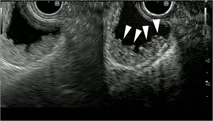
Gallbladder tumors are neoplastic lesions; however, it can be difficult to distinguish between benign and malignant gall bladder tumors before surgery, although endoscopic ultrasound (EUS) is useful for differentiation. Fundamental B mode EUS (FB-EUS) and contrast-enhanced harmonic EUS (CH-EUS) are reported to be useful for the diagnosis of gallbladder tumor because they allow evaluation of polypoid lesion and gallbladder wall thickening. Scoring systems based on FB-EUS imaging are available for the diagnosis of malignant gallbladder polypoid lesions. The characteristic findings of malignant gallbladder polypoid lesions on CH-EUS include the presence of irregular intratumoral vessels and perfusion defects. The characteristic findings of malignant gallbladder wall thickening on FB-EUS include wall thickening >12 mm, hypoechoic internal echogenicity, inhomogeneous internal echo pattern, and disrupted wall layer, whereas CH-EUS findings include hypovascular enhancement and inhomogeneous contrast distribution pattern. In addition, FB-EUS and CH-EUS are useful for evaluating the stage of gallbladder carcinoma because they allow the evaluation of the depth of invasion of the gallbladder wall. It is usually difficult to obtain pathological evidence from gallbladder tumors before surgery and chemotherapy, even though the histological diagnosis is necessary for determining treatment policy. EUS-guided fine needle aspiration (EUS-FNA) is useful for obtaining pathological samples from gallbladder tumors before surgery and chemotherapy. The accuracy rate of EUS-FNA for gallbladder tumor is as high as 90%, but complications such as bile leakage and needle track seeding can be a problem, although it was reported that contrast-enhanced harmonic imaging is useful for avoiding them.

Keywords: contrast-enhanced harmonic endoscopic ultrasonography (CH-EUS); endoscopic ultrasound (EUS); endoscopic ultrasound-guided fine needle aspiration (EUS-FNA); gallbladder carcinoma (GBC); gallbladder tumor.


Conflict of interest statement

The authors declare that the research was conducted in the absence of any commercial or financial relationships that could be construed as a potential conflict of interest.

Figures

Figure 1
Figure 1
Fundamental B-mode EUS for gallbladder polypoid lesions. Gallbladder adenoma: iso-echoic homogenous pedunculated mass lesion.
Figure 2
Figure 2
Fundamental B-mode EUS for gallbladder polypoid lesions. Gallbladder adenocarcinoma: internal hypoechoic heterogenous papillary elevated mass lesion (arrow).
Figure 3
Figure 3
Contrast-enhanced harmonic endoscopic ultrasound (EUS) for gallbladder polypoid lesions. Gallbladder adenoma: The mass is a polypoid lesion with homogeneous hyperenhancement (arrow).
Figure 4
Figure 4
Contrast-enhanced harmonic endoscopic ultrasound (EUS) for gallbladder polypoid lesions. Gallbladder adenocarcinoma: The polypoid lesion has a perfusion defect area (arrow).
Figure 5
Figure 5
Fundamental B-mode EUS for gallbladder wall thickening. Adenomyomatosis: gallbladder wall thickening (white arrow) with a uniform surface and intramural microcystic anechoic area.
Figure 6
Figure 6
Fundamental B-mode EUS for gallbladder wall thickening. Gallbladder adenocarcinoma: gallbladder wall thickening of more than 10 mm, hypoechoic internal echogenicity, inhomogeneous internal echo pattern, and disrupted wall layer.
Figure 7
Figure 7
Contrast-enhanced harmonic EUS for gallbladder wall thickening gallbladder wall thickening (arrowhead) demonstrates heterogeneous enhancement.
Figure 8
Figure 8
Fundamental B-mode EUS for staging of gallbladder carcinoma. T3 gallbladder carcinoma: hypoechoic tumor (arrow) in the gallbladder without a disrupted hyperechoic layer (arrowhead).
Figure 9
Figure 9
Fundamental B-mode EUS for staging of gallbladder carcinoma. T4 gallbladder carcinoma: heterogeneous hypoechoic tumor (arrow) in the gallbladder without a disrupted hyperechoic layer (arrowhead).
Figure 10
Figure 10
Contrast-enhanced harmonic EUS for staging of gallbladder carcinoma. T3 gallbladder carcinoma: heterogenous hyperenhancing tumor (arrow) in the gallbladder without disrupted hyperenhancement in the outer layer (arrowhead).
Figure 11
Figure 11
Contrast-enhanced harmonic EUS for staging of gallbladder carcinoma. T4 gallbladder carcinoma: heterogenous hypoenhancing tumor (arrow) in the gallbladder with disrupted hyperenhacement in the outer layer (arrowhead).

Similar articles

Cited by

References

    1. Chantarojanasiri T, Hirooka Y, Kawashima H, Ohno E, Kongkam P, Goto H. The role of endoscopic ultrasound in the diagnosis of gallbladder diseases. J Med Ultrason. (2017) 44:63–70. 10.1007/s10396-016-0742-9 - DOI - PubMed
    1. Hijioka S, Nagashio Y, Ohba A, Maruki Y, Okusaka T. The role of EUS and EUS-FNA in differentiating benign and malignant gallbladder lesions. Diagnostics. (2021) 11:1586. 10.3390/diagnostics11091586 - DOI - PMC - PubMed
    1. Kamata K, Takenaka M, Kitano M, Omoto S, Miyata T, Minaga K, et al. . Contrast-enhanced harmonic endoscopic ultrasonography for differential diagnosis of localized gallbladder lesions. Dig Endosc Off J Jpn Gastroenterol Endosc Soc. (2018) 30:98–106. 10.1111/den.12900 - DOI - PubMed
    1. Hijioka S, Hara K, Mizuno N, Imaoka H, Ogura T, Haba S, et al. . Diagnostic yield of endoscopic retrograde cholangiography and of EUS-guided fine needle aspiration sampling in gallbladder carcinomas. J Hepatobil Pancreat Sci. (2012) 19:650–5. 10.1007/s00534-011-0482-6 - DOI - PubMed
    1. Hijioka S, Mekky MA, Bhatia V, Sawaki A, Mizuno N, Hara K, et al. . Can EUS-guided FNA distinguish between gallbladder cancer and xanthogranulomatous cholecystitis? Gastrointest Endosc. (2010) 72:622–7. 10.1016/j.gie.2010.05.022 - DOI - PubMed

LinkOut - more resources