. 2023:2564:185-201.
doi: 10.1007/978-1-0716-2667-2_8.

CRISPR-Cas9-Mediated Knock-In Approach to Insert the GFP11 Tag into the Genome of a Human Cell Line

Affiliations

CRISPR-Cas9-Mediated Knock-In Approach to Insert the GFP11 Tag into the Genome of a Human Cell Line

Ryo Tamura et al. Methods Mol Biol. 2023.

Abstract

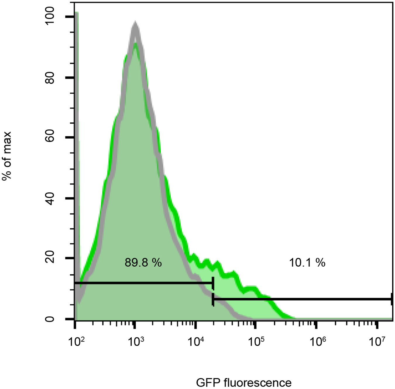
The protocol in this chapter describes a method to label endogenous proteins using a self-complementing split green fluorescent protein (split GFP1-10/11) in a human cell line. By directly delivering Cas9/sgRNA ribonucleoprotein (RNP) complexes through nucleofection, this protocol allows for the efficient integration of GFP11 into a specific genomic locus via CRISPR-Cas9-mediated homology-directed repair (HDR). We use the GFP11 sequence in the form of a single-stranded DNA (ssDNA) as an HDR template. Because the ssDNA with less than 200 nucleotides used here is commercially synthesized, this approach remains cloning-free. The integration of GFP11 is performed in cells stably expressing GFP1-10, thereby inducing fluorescence reconstitution. Subsequently, such a reconstituted signal is analyzed using fluorescence flow cytometry for estimating knock-in efficiencies and enriching the GFP-positive cell population. Finally, the enriched cells can be visualized using fluorescence microscopy.

Keywords: CRISPR; Cas9; GFP1-10; GFP11; Homology-directed repair; Split GFP.


Figures

Figure 1.
Figure 1.
Overview of GFP11 knock-in generation via CRISPR-Cas9-mediated HDR. Synthetic oligonucleotide primers are used to generate a DNA template. The DNA template enables the production of sgRNA through in vitro transcription. A Cas9/sgRNA ribonucleoprotein (RNP) complex is assembled in vitro. An ssDNA HDR repair template and the Cas9/sgRNA RNP complex are nucleofected into HEK 293 cells. GFP11 knock-in cells can be analyzed by flow cytometry to assess the knock-in efficiency and enrich GFP-positive cells or be imaged by fluorescence microscopy to localize the tagged proteins.
Figure 2.
Figure 2.
Example of an HDR repair template design. Top: An HDR template contains a 3-aa linker (black bold upper case), a GFP11 coding sequence (green bold upper case), and two flanking homology arms for recombination (black lower case). The template is designed to integrate GFP11 to the 3’ end of the HIST2H2BE gene immediately upstream of the stop codon (underlined lower case). Bottom: A region within the HIST2H2BE locus harboring a ~20 nucleotide sequence for the sgRNA site (purple underlined lower case).
Figure 3.
Figure 3.
Example of a sgRNA design. (a) A DNA template eventually used for in vitro transcription of sgRNA (Step 5 in the 3.1.2 section). This template holds a T7 promoter, a gene-specific sequence, and a common sgRNA scaffold. (b) An overlapping PCR scheme for the synthesis of the DNA template. The DNA template is PCR-amplified using three common primers (T25, ML611, BS7) and a gene-specific primer. See Table 1 for the 5’ and 3’ primer sequences.
Figure 4.
Figure 4.
A representative flow cytometry histogram of GFP11 knock-in HEK 293 cells (green). To estimate the GFP11 knock-in efficiency a the HIST2H2BE locus, GFP1-10-expressing cells were used as a non-nucleofected control (gray). In this example, ~ 10% of the nucleofected cells are GFP-positive.
Figure 5.
Figure 5.
A representative confocal image of GFP11 knock-in HEK 293 cells. Endogenous H2B proteins are labeled with split GFP. The resulting reconstituted GFP signal displays the nuclear localization of Histone 2B. Scale bar, 20 μm.

References

    1. Leroy H, Lee R (2013) The Human Genome Project: big science transforms biology and medicine. Genome Medicine 5, 79. - PMC - PubMed
    1. Shaner NC, Patterson GH, Davidson MW (2007) Advances in fluorescent protein technology. J Cell Sci 120, 4247–4260. - PubMed
    1. Rizzo MA, Davidson MW, Piston DW (2009) Fluorescent protein tracking and detection: applications using fluorescent proteins in living cells. Cold Spring Harb Protoc 2009. - PubMed
    1. Lu W, Lakonishok M, Gelfand VI (2021) Gatekeeper function for Short stop at the ring canals of the Drosophila ovary. Curr Biol 31, 3207–3220. - PMC - PubMed
    1. Sengupta P, Seo AY, Pasolli HA, Song YE, Johnson MC, Lippincott-Schwartz J (2019) A lipid-based partitioning mechanism for selective incorporation of proteins into membranes of HIV particles. Nat Cell Biol 21, 452–461. - PubMed

Publication types

LinkOut - more resources