. 2022 Sep 23;50(17):9689-9704.
doi: 10.1093/nar/gkac739.

Global 5'-UTR RNA structure regulates translation of a SERPINA1 mRNA

Affiliations

Global 5'-UTR RNA structure regulates translation of a SERPINA1 mRNA

Philip J Grayeski et al. Nucleic Acids Res. .

Abstract

SERPINA1 mRNAs encode the protease inhibitor α-1-antitrypsin and are regulated through post-transcriptional mechanisms. α-1-antitrypsin deficiency leads to chronic obstructive pulmonary disease (COPD) and liver cirrhosis, and specific variants in the 5'-untranslated region (5'-UTR) are associated with COPD. The NM_000295.4 transcript is well expressed and translated in lung and blood and features an extended 5'-UTR that does not contain a competing upstream open reading frame (uORF). We show that the 5'-UTR of NM_000295.4 folds into a well-defined multi-helix structural domain. We systematically destabilized mRNA structure across the NM_000295.4 5'-UTR, and measured changes in (SHAPE quantified) RNA structure and cap-dependent translation relative to a native-sequence reporter. Surprisingly, despite destabilizing local RNA structure, most mutations either had no effect on or decreased translation. Most structure-destabilizing mutations retained native, global 5'-UTR structure. However, those mutations that disrupted the helix that anchors the 5'-UTR domain yielded three groups of non-native structures. Two of these non-native structure groups refolded to create a stable helix near the translation initiation site that decreases translation. Thus, in contrast to the conventional model that RNA structure in 5'-UTRs primarily inhibits translation, complex folding of the NM_000295.4 5'-UTR creates a translation-optimized message by promoting accessibility at the translation initiation site.


Figures

Figure 1.
Figure 1.
Strategy for analysis of structure-function relationships across a 5′-UTR. Structure-destabilizing substitutions (5′-UUAUUA-3′) were tiled across the SERPINA1 NM_000295.4 5′-UTR region in a luciferase reporter mRNA. Structural effects of mutation were assessed by nucleotide-resolution chemical probing (SHAPE-MaP) and data-directed structural modeling. Functional effects were evaluated in reporter translation assays.
Figure 2.
Figure 2.
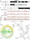
Structure of the NM_000295.4 5′-UTR and CDS. (A) Structures of the pre-mRNA of SERPINA1 isoform NM_000295.4 and plasmid reporter. The reporter mRNA contains the entire spliced 5′-UTR and 240 nucleotides of the CDS, inserted downstream of a CMV promoter (45 nucleotides). Arrows indicate primers used to selectively analyze endogenous and plasmid-based mRNAs. (B) SHAPE profiles for the endogenous NM_000295.4 mRNA expressed in hepatocytes (HepG2), and for the native sequence reporter construct, expressed in HEK293T cells. Data for cell-free and in-cell probing are shown. The transcription start site for NM_000295.4 is annotated as +1. Short gray bars indicate nucleotides with high inter-replicate variability (>50%) for in-cell experiment with endogenous RNA. (C) Arc diagrams showing pairing probabilities for base pairs modeled under cell-free and in-cell conditions for the native sequence NM_000295.4 5′-UTR and CDS, encoded by the reporter plasmid. Pairing probabilities are indicated by color scale. (D) Secondary structure model for native sequence NM_000295.4 5′-UTR and CDS (240 nucleotides) under cell-free conditions. Nucleotides are colored by SHAPE reactivity. The overall structure is conserved between endogenous and plasmid-encoded RNAs under both cell-free and in-cell conditions (Supplementary Figure S2, Tables S1 and S2).
Figure 3.
Figure 3.
Consequences of local structure-destabilizing mutations across the NM_000295.4 5′-UTR and initial CDS. (A) Heat map of SHAPE reactivity changes—quantified as ΔSHAPE changes (44)—for each mutant relative to the native sequence transcript. Mutation sites are indicated by black bars; mutants are named by the position of their 3′-most substituted nucleotide. Increases and decreases are shown on a red to blue scale. (B) Arc diagrams, linear pairing probability plots, and ΔSHAPE (middle) for representative structure-altering mutant (mutant 90) versus native sequence transcript. Site of mutation indicated by black bar.
Figure 4.
Figure 4.
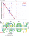
5′-UTR mutations form RNA structures that cluster into distinct groups. (A) Linear pairing probabilities for the native sequence and 42 mutant 5′-UTRs. A principal component analysis, based on similarity in pairing probabilities, yielded four clusters, native-like and groups 1, 2 and 3; groupings were supported by k-means clustering. Features, indicated by numbers below plots, denote regions with characteristic structural differences between groups. (B) Arc diagrams showing pairing probabilities for the native sequence structure and a representative mutant from each structure group. Sensitivity (sens) and positive predictive value (ppv) calculated from pairing probabilities (≥0.1 threshold) as compared to the native sequence 5′-UTR structure.
Figure 5.
Figure 5.
Translation depends on NM_000295.4 structure. (A) Translation of mutants relative to the native sequence construct, measured by dual luciferase assay. Mutants are ordered 5′ to 3′ and colored by structural group classification. Error bars show standard deviations (n = 6, two plasmid replicates each comprising three biological replicates). (B) Distribution of relative translation, as a function of structural group. Individual mutants are plotted as points; native sequence construct is shown in red. Median is shown as horizontal line; boxes show the interquartile range [IQR, from quartile 1 (Q1) to quartile 3 (Q3)]; whiskers highlight the range, Q1 – 1.5 × IQR to Q3 + 1.5 × IQR. *P ≤ 0.05 (two-tailed t-test). (C) Superposition of mutation positions that folded into non-native global structures and altered translation by ≥15% on the native sequence NM_000295.4 5′-UTR structure (based on cell-free data).
Figure 6.
Figure 6.
Translation is anti-correlated with structure at the translation initiation site. Relationship between translation and energetic cost of unfolding structures at the translation initiation site for each structure group. ΔGunfold was calculated for a window of ±15 nucleotides from the adenosine of the start codon (analysis of alternative window sizes shown in Supplementary Figure S4). Relationship between translation and cost of structure unfolding (ΔGunfold) is specific to the translation initiation site (Supplementary Figure S6). Data are shown as two-dimensional box plots; in both dimensions, boxes span the IQR; whiskers extend to minimum and maximum observed values. Individual mutants are plotted as points; the native sequence is red. *P ≤ 0.05; **P≤ 0.001 (two-tailed t-test).
Figure 7.
Figure 7.
Model for regulation of translation by NM_000295.4 5′-UTR structure. Comparison of Stem 1 in the native sequence with alternative structures formed in each mutant structure group. Local RNA structure changes near the translation start site (shaded box) highlighted. Asterisks denote well-defined hairpins formed in group 1 and 2 mutants. ΔGunfold calculated for the 30 nucleotide window emphasized with shaded box.

References

    1. Brantly M., Nukiwa T., Crystal R.G.. Molecular basis of alpha-1-antitrypsin deficiency. Am. J. Med. 1988; 84:13–31. - PubMed
    1. Brantly M. α1-antitrypsin: not just an antiprotease: extending the half-life of a natural anti-inflammatory molecule by conjugation with polyethylene glycol. Am. J. Respir. Cell Mol. Biol. 2002; 27:652–654. - PubMed
    1. Carlson J.A., Rogers B.B., Sifers R.N., Hawkins H.K., Finegold M.J., Woo S.L.C.. Multiple tissues express alpha 1-antitrypsin in transgenic mice and man. J. Clin. Invest. 1988; 82:26–36. - PMC - PubMed
    1. Corley M., Solem A., Phillips G., Lackey L., Ziehr B., Vincent H.A., Mustoe A.M., Ramos S.B.V, Weeks K.M., Moorman N.J.et al. .. An RNA structure-mediated, posttranscriptional model of human α-1-antitrypsin expression. Proc. Natl. Acad. Sci. U.S.A. 2017; 114:E10244–E10253. - PMC - PubMed
    1. Lackey L., McArthur E., Laederach A.. Increased transcript complexity in genes associated with chronic obstructive pulmonary disease. PLoS One. 2015; 10:e0140885. - PMC - PubMed

Publication types

MeSH terms