. 2022 Sep 23;50(17):9893-9908.
doi: 10.1093/nar/gkac745.

Tracking of progressing human DNA polymerase δ holoenzymes reveals distributions of DNA lesion bypass activities

Affiliations

Tracking of progressing human DNA polymerase δ holoenzymes reveals distributions of DNA lesion bypass activities

Rachel L Dannenberg et al. Nucleic Acids Res. .

Abstract

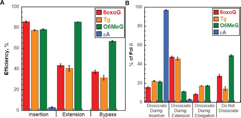
During DNA replication, DNA lesions in lagging strand templates are initially encountered by DNA polymerase δ (pol δ) holoenzymes comprised of pol δ and the PCNA processivity sliding clamp. These encounters are thought to stall replication of an afflicted template before the lesion, activating DNA damage tolerance (DDT) pathways that replicate the lesion and adjacent DNA sequence, allowing pol δ to resume. However, qualitative studies observed that human pol δ can replicate various DNA lesions, albeit with unknown proficiencies, which raises issues regarding the role of DDT in replicating DNA lesions. To address these issues, we re-constituted human lagging strand replication to quantitatively characterize initial encounters of pol δ holoenzymes with DNA lesions. The results indicate pol δ holoenzymes support dNTP incorporation opposite and beyond multiple lesions and the extent of these activities depends on the lesion and pol δ proofreading. Furthermore, after encountering a given DNA lesion, subsequent dissociation of pol δ is distributed around the lesion and a portion does not dissociate. The distributions of these events are dependent on the lesion and pol δ proofreading. Collectively, these results reveal complexity and heterogeneity in the replication of lagging strand DNA lesions, significantly advancing our understanding of human DDT.


Figures

Figure 1.
Figure 1.
DNA damage tolerance in lagging strand templates. At the top, a progressing pol δ holoenzyme (pol δ + PCNA) is depicted replicating a lagging strand template engaged by RPA. 1) A progressing pol δ holoenzyme encounters a DNA lesion in a lagging strand template. 2) Pol δ rapidly and passively dissociates into solution, leaving PCNA and RPA behind on the DNA. Pol δ may reiteratively associate and dissociate to/from the resident PCNA encircling the stalled P/T junction but cannot support stable dNTP incorporation opposite the offending DNA lesion. 3) The stalled P/T junction activates one or more DNA damage tolerance pathway(s) that are ultimately responsible for the insertion of a dNTP opposite the lesion (insertion), extension of the nascent DNA 1 nt downstream of the lesion (extension), and possibly further extension of the nascent DNA > 1 nt downstream of the lesion (elongation). TLS is depicted as an example of DDT. In this pathway, one or more TLS polymerases engage PCNA encircling the aborted P/T junction and perform insertion, extension, and (possibly) elongation. 4) After DDT is complete, replication by a pol δ holoenzyme may resume downstream of the lesion. In this view, DDT is solely responsible for the replication of a DNA lesion (i.e. insertion), and, hence, pol δ does not contribute to the fidelity of replicating DNA lesions.
Figure 2.
Figure 2.
Replication by pol δ holoenzymes. (A) Schematic representation of the experiment performed to monitor primer extension by pol δ holoenzymes during a single binding encounter with a P/T DNA substrate. (B) 16% denaturing sequencing gel of the primer extension products observed for the native (i.e. undamaged) DNA substrate (BioCy5P/T, Supplementary Figure S1). The incorporation step (i) for certain primer extension products (i1 to i10, i12 and i33) is indicated on the far right. Shown on the left and the right are representative gels of primer extension by pol δ observed in the absence (‘–PCNA’) and presence (‘+ PCNA’) of PCNA, respectively. (C) Quantification of the (total) primer extension products. Data is plotted as a function of time (after the addition of pol δ) and display ‘burst’ kinetics. Data points within the ‘linear’ phase are fit to a linear regression where the Y-intercept (in nM) represents the amplitude of the ‘burst’ phase, and the slope represents the initial velocity (in nM/s) of the linear phase. Data for experiments carried out in the absence (‘–PCNA’) and presence (‘+PCNA’) of PCNA are displayed in grey and black, respectively. Data for experiments carried out in the presence of PCNA are fit to a burst + linear phase kinetic model (dashed line) only for visualizing the conformity of the linear phases for each fit. (D) Processivity of pol δ holoenzymes. The probability of incorporation (Pi) for each dNTP incorporation step (i) beyond the first incorporation step is calculated as described in Materials and Methods. Pi values observed in the presence of PCNA are plotted as a function of the dNTP incorporation step i. Data is fit to an interpolation only for observation.
Figure 3.
Figure 3.
Pol δ holoenzymes encountering an 8oxoG lesion downstream of a P/T junction. The progression of human pol δ holoenzymes was monitored on a P/T DNA substrate (Bio-Cy5-P/T-8oxoG, Supplementary Figure S1) that contains an 8oxoG 12 nt downstream of the P/T junction (at the 12th dNTP incorporation step, i12). (A) Structure of 8oxoG. 8oxoG (Bottom) is generated from G (top) through the introduction of an oxo group on the carbon at position 8 and the addition of a hydrogen to the nitrogen at position 7. These modifications are highlighted in red on the structure of 8oxoG. (B) 16% denaturing sequencing gel of the primer extension products. Shown on the left and the right are representative gels of primer extension by pol δ holoenzymes on the native Bio-Cy5-P/T (‘G at i12’) and the Bio-Cy5-P/T-8oxoG (‘8oxoG at i12’) DNA substrates, respectively. (C) Processivity of pol δ holoenzymes. Pi values observed for the native Bio-Cy5-P/T (‘G at i12’) and the Bio-Cy5-P/T-8oxoG (‘8oxoG at i12’) DNA substrates are shown in black and red, respectively, and plotted as a function of the dNTP incorporation step i. Data is fit to an interpolation only for observation. Dashed line indicates dNTP incorporation step for insertion (i12). (D) Efficiency of replicating 8oxoG. The efficiencies for insertion, extension, and bypass are calculated as described in Materials and Methods and plotted as percentages. The Pi value(s) from which each efficiency is derived from is indicated below the respective efficiency. Values for each parameter are also reported in Supplementary Table S1. (E). Dissociation of pol δ holoenzymes after encountering an 8oxoG lesion at i12. The distribution of pol δ dissociation events observed for the native Bio-Cy5-P/T (‘G at i12’) and the Bio-Cy5-P/T-8oxoG (‘8oxoG at i12’) DNA substrates are indicated in black and red, respectively.
Figure 4.
Figure 4.
Effect of proofreading on bypass of 8oxoG by pol δ holoenzymes. (A) Efficiency of replicating 8oxoG. The efficiencies for insertion, extension, and bypass for wild-type (WT) and exonuclease-deficient (Exo–) pol δ holoenzymes are plotted as percentages. Values for each parameter are also reported in Supplementary Table S1. (B) Dissociation of pol δ holoenzymes after encountering an 8oxoG lesion at i12. The distribution of dissociation events observed for the Bio-Cy5-P/T-8oxoG DNA substrate with wild-type (WT) and exonuclease-deficient (Exo–) pol δ holoenzymes are plotted.
Figure 5.
Figure 5.
Pol δ holoenzymes encountering a Tg lesion downstream of a P/T junction. The progression of human pol δ holoenzymes was monitored on a P/T DNA substrate (Bio-Cy5-P/T-Tg, Supplementary Figure S1) that contains a Tg 9 nt downstream of the P/T junction (at the 9th dNTP incorporation step, i9). (A) Structure of Tg. Tg (bottom) is generated from T (top) through the addition of hydroxyl groups on the carbons at position 5 and position 6 of the ring. This results in a loss of aromaticity and conversion from planar to nonplanar. These modifications are highlighted in orange on the structure of Tg. (B) 16% denaturing sequencing gel of the primer extension products. Shown on the left and the right are representative gels of primer extension by pol δ holoenzymes on the native Bio-Cy5-P/T (‘T at i9’) and the Bio-Cy5-P/T-Tg (‘Tg at i9’) DNA substrates, respectively. (C) Processivity of pol δ holoenzymes. Pi values observed for the native Bio-Cy5-P/T (‘T at i9’) and the Bio-Cy5-P/T-Tg (‘Tg at i9’) DNA substrates are shown in black and orange, respectively, and plotted as a function of the dNTP incorporation step, i. Data is fit to an interpolation only for observation. Dashed line indicates dNTP incorporation step for insertion (i9). (D) Efficiency of replicating Tg. The efficiencies for insertion, extension, and bypass are calculated as described in Materials and Methods and plotted as percentages. Values for each parameter are also reported in Supplementary Table S2. (E). Dissociation of pol δ holoenzymes after encountering a Tg lesion. The distribution of dissociation events observed for the native Bio-Cy5-P/T (‘T at i9’) and the Bio-Cy5-P/T-Tg (‘Tg at i9’) DNA substrates are indicated by black and orange, respectively.
Figure 6.
Figure 6.
Effect of proofreading on bypass of Tg by pol δ holoenzymes. (A) Efficiency of replicating Tg. The efficiencies for insertion, extension, and bypass for wild-type (WT) and exonuclease-deficient (Exo–) pol δ holoenzymes are plotted as percentages. Values for each parameter are also reported in Supplementary Table S2. (B) Dissociation of pol δ holoenzymes after encountering an Tg lesion. The distribution of dissociation events observed for the Bio-Cy5-P/T-Tg DNA substrate with wild-type (WT) and exonuclease-deficient (Exo–) pol δ holoenzymes are plotted.
Figure 7.
Figure 7.
Pol δ holoenzymes encountering an O6MeG lesion downstream of a P/T junction. The progression of human pol δ holoenzymes was monitored on a P/T DNA substrate (Bio-Cy5-P/T-O6MeG, Supplementary Figure S1) that contains an O6MeG 12 nt downstream of the P/T junction (at the 12th dNTP incorporation step, i12). (A) Structure of O6MeG. O6MeG (bottom) is generated from G (top) through the addition of a methyl group on the oxygen of the carbonyl group at position 6 of the ring. These modifications are highlighted in green on the structure of O6MeG. (B) 16% denaturing sequencing gel of the primer extension products. Shown on the left and the right are representative gels of primer extension by pol δ holoenzymes on the native Bio-Cy5-P/T (‘G at i12’) and the Bio-Cy5-P/T-O6MeG (‘O6MeG at i12’) DNA substrates, respectively. (C) Processivity of pol δ holoenzymes. Pi values observed for the native Bio-Cy5-P/T (‘G at i12’) and the Bio-Cy5-P/T-O6MeG (‘O6MeG at i12’) DNA substrates are shown in black and green, respectively, and plotted as a function of the dNTP incorporation step, i. Data is fit to an interpolation only for observation. Dashed line indicates dNTP incorporation step for insertion (i12). (D) Efficiency of replicating O6MeG. The efficiencies for insertion, extension, and bypass are calculated as described in Materials and Methods and plotted as percentages. Values for each parameter are also reported in Supplementary Table S3. (E). Dissociation of pol δ holoenzymes after encountering an O6MeG lesion. The distribution of dissociation events observed for the native Bio-Cy5-P/T (‘G at i12’) and the Bio-Cy5-P/T-O6MeG (‘O6MeG at i12’) DNA substrates are indicated by black and green, respectively.
Figure 8.
Figure 8.
Effect of proofreading on bypass of O6MeG by pol δ holoenzymes. (A) Efficiency of replicating O6MeG. The efficiencies for insertion, extension and bypass for wild-type (WT) and exonuclease-deficient (Exo–) pol δ holoenzymes are plotted as percentages. Values for each parameter are also reported in Supplementary Table S3. (B) Dissociation of pol δ holoenzymes after encountering an O6MeG lesion. The distribution of dissociation events observed for the Bio-Cy5-P/T-O6MeG DNA substrate with wild-type (WT) and exonuclease-deficient (Exo–) pol δ holoenzymes are plotted.
Figure 9.
Figure 9.
Pol δ holoenzymes encountering an ϵA lesion downstream of a P/T junction. The progression of human pol δ holoenzymes was monitored on a P/T DNA substrate (Bio-Cy5-P/T-ϵA, Supplementary Figure S1) that contains an ϵA 10 nt downstream of the P/T junction (at the 10th dNTP incorporation step, i10). (A) Structure of ϵA. ϵA (bottom) is generated from A (top) through the attachment of two extra carbons in an exocyclic arrangement; 1 carbon is attached to the nitrogen at position 1 and the other is attached to the nitrogen in the amine at position 6 of the ring. These modifications are highlighted in blue on the structure of ϵA. (B) 16% denaturing sequencing gel of the primer extension products. Shown on the left and the right are representative gels of primer extension by pol δ holoenzymes on the native Bio-Cy5-P/T (‘A at i10’) and the Bio-Cy5-P/T-ϵA (‘ϵA at i10’) DNA substrates, respectively. (C) Processivity of pol δ holoenzymes. Pi values observed for the native Bio-Cy5-P/T (‘A at i10’) and the Bio-Cy5-P/T-ϵA (‘ϵA at i10’) DNA substrates are shown in black and blue, respectively, and plotted as a function of the dNTP incorporation step, i. Data is fit to an interpolation only for observation. (D) Efficiency of replicating ϵA. The efficiencies for insertion, extension and bypass are calculated as described in Materials and Methods and plotted as percentages. Values for each parameter are also reported in Supplementary Table S4. (E). Dissociation of pol δ holoenzymes after encountering an ϵA lesion. The distribution of dissociation events observed for the native Bio-Cy5-P/T (‘A at i10’) and the Bio-Cy5-P/T-ϵA (‘ϵA at i10’) DNA substrates are indicated by black and blue, respectively.
Figure 10.
Figure 10.
Effect of proofreading on bypass of ϵA by pol δ holoenzymes. (A) Efficiency of replicating ϵA. The efficiencies for insertion, extension, and bypass for wild-type (WT) and exonuclease-deficient (Exo-) pol δ holoenzymes are plotted as percentages. Values for each parameter are also reported in Supplementary Table S4. (B) Dissociation of pol δ holoenzymes after encountering an ϵA lesion. The distribution of dissociation events observed for the Bio-Cy5-P/T-ϵA DNA substrate with wild-type (WT) and exonuclease-deficient (Exo-) pol δ holoenzymes are plotted.
Figure 11.
Figure 11.
Bypass of DNA lesions by pol δ holoenzymes during initial encounters. (A) Efficiencies of replicating DNA lesions. The efficiencies for dNTP incorporation opposite a lesion (i.e. insertion), 1 nt downstream of lesion (i.e. extension) and bypass (insertion and extension) of a lesion for wild type pol δ holoenzymes are plotted. Data is taken from Figures 3D, 5D, 7D and 9D and is color-coded by DNA lesion. (B) Dissociation of pol δ holoenzymes after encountering DNA lesions. The distribution of dissociation events observed for wild type pol δ holoenzymes encountering DNA lesions is plotted. Data is taken from Figures 3E, 5E, 7E and 9E and is color-coded by DNA lesion.

Similar articles

Cited by

References

    1. Lee M., Wang X., Zhang S., Zhang Z., Lee E.Y.C.. Regulation and modulation of human DNA polymerase delta activity and function. Genes (Basel). 2017; 8:190. - PMC - PubMed
    1. Hedglin M., Pandey B., Benkovic S.J.. Stability of the human polymerase delta holoenzyme and its implications in lagging strand DNA synthesis. Proc. Natl. Acad. Sci. U.S.A. 2016; 113:E1777–E1786. - PMC - PubMed
    1. Hedglin M., Kumar R., Benkovic S.J.. Replication clamps and clamp loaders. Cold Spring Harb. Perspect. Biol. 2013; 5:a010165. - PMC - PubMed
    1. Chen R., Wold M.S.. Replication protein A: single-stranded DNA’s first responder: dynamic DNA-interactions allow replication protein a to direct single-strand DNA intermediates into different pathways for synthesis or repair. Bioessays. 2014; 36:1156–1161. - PMC - PubMed
    1. Hedglin M., Aitha M., Benkovic S.J.. Monitoring the retention of human proliferating cell nuclear antigen at primer/template junctions by proteins that bind single-stranded DNA. Biochemistry. 2017; 56:3415–3421. - PMC - PubMed

Publication types