. 2022 Sep 15;17(9):e0274420.
doi: 10.1371/journal.pone.0274420. eCollection 2022.

UDP-glucose dehydrogenase (UGDH) activity is suppressed by peroxide and promoted by PDGF in fibroblast-like synoviocytes: Evidence of a redox control mechanism

Affiliations

UDP-glucose dehydrogenase (UGDH) activity is suppressed by peroxide and promoted by PDGF in fibroblast-like synoviocytes: Evidence of a redox control mechanism

Ramya Chandrasekaran et al. PLoS One. .

Abstract

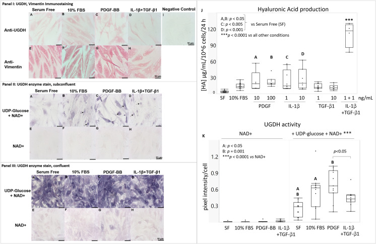
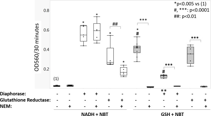
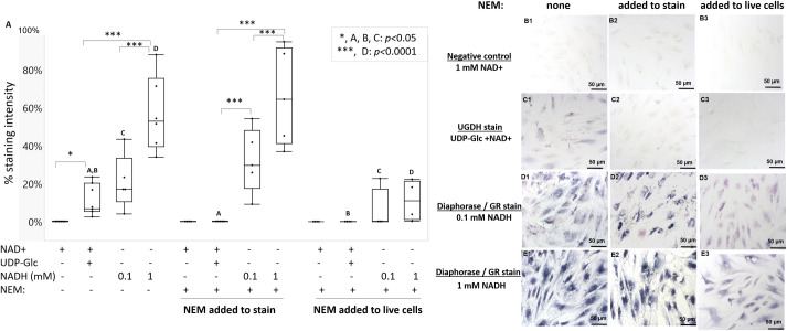
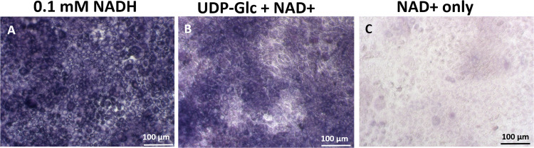
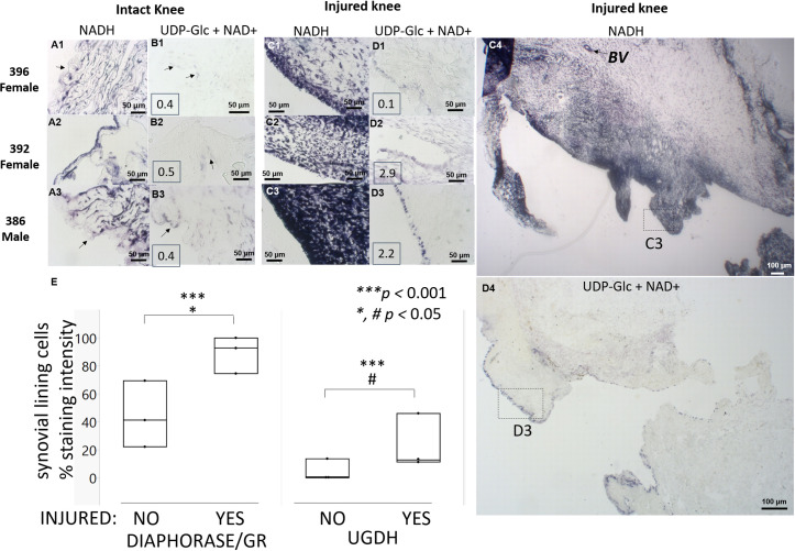
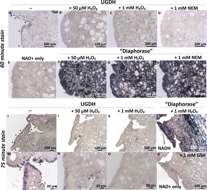
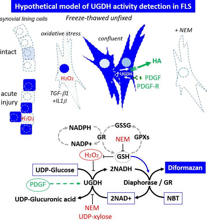
UDP-glucose dehydrogenase (UGDH) generates essential precursors of hyaluronic acid (HA) synthesis, however mechanisms regulating its activity are unclear. We used enzyme histostaining and quantitative image analysis to test whether cytokines that stimulate HA synthesis upregulate UGDH activity. Fibroblast-like synoviocytes (FLS, from N = 6 human donors with knee pain) were cultured, freeze-thawed, and incubated for 1 hour with UDP-glucose, NAD+ and nitroblue tetrazolium (NBT) which allows UGDH to generate NADH, and NADH to reduce NBT to a blue stain. Compared to serum-free medium, FLS treated with PDGF showed 3-fold higher UGDH activity and 6-fold higher HA release, but IL-1beta/TGF-beta1 induced 27-fold higher HA release without enhancing UGDH activity. In selected proliferating cells, UGDH activity was lost in the cytosol, but preserved in the nucleus. Cell-free assays led us to discover that diaphorase, a cytosolic enzyme, or glutathione reductase, a nuclear enzyme, was necessary and sufficient for NADH to reduce NBT to a blue formazan dye in a 1-hour timeframe. Primary synovial fibroblasts and transformed A549 fibroblasts showed constitutive diaphorase/GR staining activity that varied according to supplied NADH levels, with relatively stronger UGDH and diaphorase activity in A549 cells. Unilateral knee injury in New Zealand White rabbits (N = 3) stimulated a coordinated increase in synovial membrane UGDH and diaphorase activity, but higher synovial fluid HA in only 2 out of 3 injured joints. UGDH activity (but not diaphorase) was abolished by N-ethyl maleimide, and inhibited by peroxide or UDP-xylose. Our results do not support the hypothesis that UGDH is a rate-liming enzyme for HA synthesis under catabolic inflammatory conditions that can oxidize and inactivate the UGDH active site cysteine. Our novel data suggest a model where UGDH activity is controlled by a redox switch, where intracellular peroxide inactivates, and high glutathione and diaphorase promote UGDH activity by maintaining the active site cysteine in a reduced state, and by recycling NAD+ from NADH.


Conflict of interest statement

I have read the journal’s policy and the authors of this manuscript have the following competing interests: CDH, Scientific Advisory Board and a shareholder of Chitogenx Inc. (formerly Ortho RTi Inc.); RC, CM, RS, AC, DF, RM, HEG, SA and MP have declared that no competing interests exist. This does not alter our adherence to PLOS ONE policies on sharing data and materials.

Figures

Fig 1
Fig 1. In vitro cultured primary human FLS express UGDH and vimentin protein, and show differential HA synthetic rates and UGDH activity in response to serum and different cytokines.
FLS monolayers were cultured overnight in (A, E) serum-free, (B, F) 10% FBS, (C, G) 100 ng/mL PDGF, or (D, H) 1 ng each IL1β + TGFβ1. (Panel I) Monolayers were fixed and analyzed by immunostaining for (A-D) UGDH protein, (E-H) vimentin protein, or (I) no primary antibody. (Panels II & III) Other monolayers were frozen unfixed then submitted to enzyme histostaining for (A-D) UGDH, and (E-H) NAD+ and NBT as negative controls; representative images show enzyme staining results in areas with (Panel II) low or (Panel III) high cell density. (J) HA synthetic rate was measured after 24 hours of culture in serum-free medium supplemented or not with serum or cytokines (N = 6 donors, 3 male, 3 female). (K) UGDH enzyme activity was measured indirectly by quantitative histomorphometry of staining intensity (N = 5 donors, 2 male, 3 female, 9–12 distinct culture wells per condition). In (E-H, and K), NAD+ without UDP-Glc is the negative control for non-specific staining. Scale bars: 10 μm. Panels J & K show the median, 25% quartile range (box), min-max (whiskers); Significant differences are indicated by letters (A, B, C, D) and ***. For panel K: PDGF was 100 ng/mL, IL1β + TGFβ1 were 1 ng/mL each; SF: serum-free.
Fig 2
Fig 2. NADH required diaphorase or GR to reduce NBT whereas GSH spontaneously reduced NBT within 30 minutes of reaction.
Cell-free assays were used to measure the OD560 of reduced NBT absorbance, for mixtures of 1 mM each NADH and NBT (white boxes) or 1 mM each GSH and NBT (grey boxes) after 30 minutes of reaction without or with enzymes or NEM (median, 25% quartile range (box) and min-max (whiskers), N = 3 to 6 independent assays per condition). Symbols: ** evidence that diaphorase can accept electrons from GSH and inhibit GSH from spontaneously reducing NADH (p<0.0001 vs no diaphorase); *, ***, #, ##: significant differences between selected conditions.
Fig 3
Fig 3. NEM abolished UGDH enzyme histostaining but only partly inhibited diaphorase staining when added to live cell cultures.
(A) Quantitative histomorphometry of staining intensity for FLS stained for NAD+ only, UGDH, or diaphorase/GR, with and without NEM. NEM was either included in the staining solution (“in stain”) or added to cell cultures at 1 mM NEM for 2 hours before enzyme staining (“added to live cells”). Representative images of histostained FLS from one female donor, showed no stain for (B1-B3) the negative control (NAD+ only), NEM-sensitive staining for (C1-C3) UGDH (UDP-Glc and NAD+), and NEM-resistant staining for (D1-D3) “low diaphorase/GR”, (E1-E3) “high diaphorase/GR stain”; (B1-E1) no NEM, (B2-E2) NEM “added to stain”, or (B3-E3) NEM “added to live cells”. In panel A, significant differences are noted with letters and symbols (*, ***). The graph in Panel (A) shows the median, 25% quartile range (box), min-max, distinct cultures with 2 female human donors and 1 male donor FLS (n = 4–7). Scale bars: 50 μm.
Fig 4
Fig 4. Intense UGDH and diaphorase enzyme histostaining activity in transformed A549 cells.
By in situ enzyme staining, A549 cells showed relatively high (A) 99.8% diaphorase/GR staining with 0.1 mM NADH, (B) 93.1% UGDH enzyme staining and (C) 12.1% background NBT staining in the absence of UDP-Glc compared to primary human FLS. Scale bars: 100 μm.
Fig 5
Fig 5. Injured rabbit knee infrapatellar synovium showed higher diaphorase/GR and UGDH activity, and enhanced synovial fluid HA levels in 2 out of 3 rabbit knees compared to contralateral intact knees.
Panels show 20x magnification images of synovial membrane from regions with maximal UGDH induction, enzyme stained in situ for diaphorase (A, C), or UGDH (B, D), in intact (A, B) and contralateral injured knees (C, D). Boxed insets in panels B1-3, D1-3 show measured synovial fluid HA levels (mg/mL). (E) Quantitative histomorphometry of % stain (median, min-max, N = 3). Panels C4, D4 show low-magnification image of male rabbit infrapatellar synovium with dotted boxes showing regions illustrated in panels C3 & D3, respectively. Symbols and abbreviations: rabbit ID and sex are shown on the upper left; arrows (A-B) are regions in intact knees where UGDH staining was detected; BV: Blood vessel. Scale bars show 50 μm or 100 μm, as indicated.
Fig 6
Fig 6. Peroxide and NEM inhibited UGDH but not diaphorase enzyme staining activity in synovial lining cells from an injured rabbit knee.
In serial synovial cryosections from a New Zealand White male rabbit knee 1 week post-microdrilling, UGDH staining (A, I, M) was inhibited by NEM (D) and by peroxide in a dose-dependent manner (B, C, J, K, N, O) but “diaphorase” was insensitive to thiol modifying inhibitors (F, G, H, L). UGDH and “diaphorase” enzyme histostaining was carried out per standard conditions, in the presence of peroxide or NEM, or with NAD+ only (±1 mM GSH) as indicated, with a 60 or 75 minute incubation at 37°C. UGDH enzyme staining was completely suppressed by NEM (D) and more suppressed by 1 mM peroxide (C, K, O) than 50 μM peroxide (B, J, N). Scale bars are 20 μm or 100 μm, as indicated.
Fig 7
Fig 7. Hypothetical model representing 2-enzyme UGDH activity staining mechanisms in cultured cells and unfixed cryosections.
Boxed molecules are enzyme staining components, encircled factors and dashed lines represent activities modulated in vivo by cytokines. Oxidative stress induced by inflammatory cytokines (TGF-β1+IL-1β) can lead to peroxide generation and reversible inactivation of UGDH unless GSH scavenges the free radicals to sustain UGDH activity. High endogenous GSH can produce background staining by directly reducing NBT and by-passing UGDH. Abbreviations: PDGF: platelet derived growth factor; GR: glutathione reductase; GSH: glutathione; H2O2: peroxide; NEM: N-ethyl maleimide (thiol alkylating agent). In the upper panel, Dark blue cells: UGDH-stained cell compartments where cytosolic diaphorase, or nuclear glutathione reductase activity is present; dashed lines: freeze-thaw permeabilized cell membranes.

Similar articles

Cited by

References

    1. Egger S, Chaikuad A, Klimacek M, Kavanagh KL, Oppermann U, Nidetzky B. Structural and kinetic evidence that catalytic reaction of human UDP-glucose 6-dehydrogenase involves covalent thiohemiacetal and thioester enzyme intermediates. J Biol Chem. 2011/11/28 ed. 2012;287: 2119–2129. doi: 10.1074/jbc.M111.313015 - DOI - PMC - PubMed
    1. Chen J, Yu Y, Gao J, Yang S. UDP-glucose Dehydrogenase: The First-step Oxidation Is an NAD+-dependent Bimolecular Nucleophilic Substitution Reaction (SN2). Int J Biol Sci. 2019;15: 341–350. doi: 10.7150/ijbs.28904 - DOI - PMC - PubMed
    1. Edwards JC. The nature and origins of synovium: experimental approaches to the study of synoviocyte differentiation. J Anat. 1994;184 (Pt 3): 493–501. - PMC - PubMed
    1. Pitsillides AA, Blake SM. Uridine diphosphoglucose dehydrogenase activity in synovial lining cells in the experimental antigen induced model of rheumatoid arthritis: an indication of synovial lining cell function. Ann Rheum Dis. 1992;51: 992–995. doi: 10.1136/ard.51.8.992 - DOI - PMC - PubMed
    1. Denk PO, Roth-Eichhorn S, Gressner AM, Knorr M. Cytokine regulation of hyaluronate production by human Tenon’s capsule fibroblasts. Current Eye Research. 2000;20: 77–80. doi: 10.1076/0271-3683(200002)2021-DFT077 - DOI - PubMed

Publication types

MeSH terms

Grants and funding

LinkOut - more resources