Review
. 2022 Sep 15;18(9):e1010770.
doi: 10.1371/journal.ppat.1010770. eCollection 2022 Sep.

Advances in understanding red blood cell modifications by Babesia

Affiliations
Review

Advances in understanding red blood cell modifications by Babesia

Hassan Hakimi et al. PLoS Pathog. .

Abstract

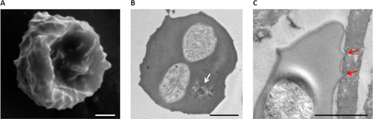
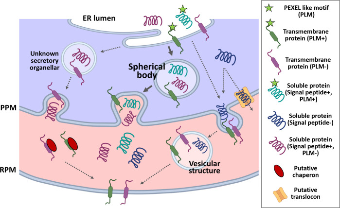
Babesia are tick-borne protozoan parasites that can infect livestock, pets, wildlife animals, and humans. In the mammalian host, they invade and multiply within red blood cells (RBCs). To support their development as obligate intracellular parasites, Babesia export numerous proteins to modify the RBC during invasion and development. Such exported proteins are likely important for parasite survival and pathogenicity and thus represent candidate drug or vaccine targets. The availability of complete genome sequences and the establishment of transfection systems for several Babesia species have aided the identification and functional characterization of exported proteins. Here, we review exported Babesia proteins; discuss their functions in the context of immune evasion, cytoadhesion, and nutrient uptake; and highlight possible future topics for research and application in this field.


Conflict of interest statement

The authors have declared that no competing interests exist.

Figures

Fig 1
Fig 1. Electron microscopy images of B. bovis-infected RBCs.
Scanning electron microscopy (A) and TEM (B) and (C) of B. bovis-iRBC binding to bovine brain endothelial cells. Membranous structures are seen in the TEM image and indicated by a white arrow. The red arrows show the ridges mediating binding of iRBCs to endothelial cells. Scale bar = 1 μm. iRBC, infected RBC; RBC, red blood cell; TEM, transmission electron microscopy.
Fig 2
Fig 2. Immunofluorescence microscopy of transgenic parasites expressing myc-tagged exported protein.
Myc-tagged proteins were episomally expressed in B. bovis (VESA1, SBP3, SmORF, BbVEAP, Bbmtm, and BbTPR-related) or B. ovata (BoMFS). The expression plasmid was constructed and transfected by electroporation as described [32,33], and transgenic parasites were selected using WR99210. Thin smears of cultured parasites were prepared for indirect immunofluorescence microscopy, fixed with a 1:1 acetone:methanol mixture, and reacted with anti-myc mouse monoclonal antibody (9B11, Cell Signaling) followed by Alexa Fluor 488-conjugated goat anti-mouse IgG (Invitrogen, green). Nuclei were stained with Hoechst 33342 (Hoechst, blue). Scale bar = 5 μm. BbVEAP, B. bovis VESA1-export associated protein; IgG, immunoglobulin G; SBP3, spherical body protein 3; SmORF, Small Open Reading Frame; VESA1, variant erythrocyte surface antigen 1.
Fig 3
Fig 3. Babesia iRBC.
Babesia parasites export numerous proteins to remodel the host RBC using secretory pathway. Several of these proteins are routed through spherical bodies, while others are either directly exported to RBC or deposited in unknown secretory organelles. B. bovis produces ridges on the surface of iRBCs to express VESA1 that mediates sequestration and immune evasion. Of spherical body proteins, SBP1, SBP2, SBP3, and SBP4 are associated with the RBC membrane; VEAP is released to the RBC cytoplasm and is essential for parasite growth and VESA1 export; mtm integrates into the RBC membrane and is likely responsible for nutrient uptake. Vesicular structures in the RBC are produced following parasite invasion that might have a role in protein export. Regarding other Babesia spp., VESA1 expression in iRBC was shown for B. orientalis and MFS export to RBC for B. ovata, though their functions remain to be determined. iRBC, infected red blood cell; mtm, multi-transmembrane protein; PPM, parasite plasma membrane; RPM, RBC plasma membrane; SBP1, spherical body protein 1; SmORF, Small Open Reading Frame protein; VEAP, VESA1-export associated protein; VESA1, variant erythrocyte surface antigen 1.
Fig 4
Fig 4. Homology clustering based on sequence similarities of ves and multi-transmembrane protein encoding genes.
(A) ves and smorf genes sequence were extracted from piroplasmaDB (https://piroplasmadb.org/piro/app), and a sequence similarity network was visualized by Gephi. (B) The figure was reproduced from Hakimi and colleagues [33]. The genes encoding proteins with more than 8 TM domains were extracted from piroplasmaDB and clustered. SmORF, Small Open Reading Frame; TM, transmembrane.
Fig 5
Fig 5. Schematic of the protein export pathway in Babesia iRBC.
The appointed soluble proteins for the export are being recruited into ER, cleaved by signal peptidase, and likely followed by cleavage of PLM. Transmembrane-containing proteins such as VESA1 or mtm are inserted into the ER membrane. These proteins are loaded into secretory vesicles of the ER-Golgi pathway, which are being transferred to spherical bodies (in the case of spherical body proteins: SBP1, SBP2, SBP3, SBP4, VEAP, and mtm) or directly to parasite plasma membrane (in the case of VESA1). Soluble proteins are released to RBC cytoplasm, while integral protein needs to be extracted from PPM, which may involve protein translocation. Soluble protein could reach the target by diffusion, while membrane proteins are carried out to the target destination through vesicles or in complex with chaperones. It is noted that this scheme is speculative. ER, endoplasmic reticulum; iRBC, infected RBC; PLM, PEXEL-like motif; PPM, parasite plasma membrane; RBC, red blood cell; RPM, RBC plasma membrane; VESA1, variant erythrocyte surface antigen 1.

References

    1. Schnittger L, Rodriguez AE, Florin-Christensen M, Morrison DA. Babesia: a world emerging. Infect Genet Evol. 2012;12(8):1788–809. Epub 20120731. doi: 10.1016/j.meegid.2012.07.004 . - DOI - PubMed
    1. Bock R, Jackson L, de Vos A, Jorgensen W. Babesiosis of cattle. Parasitology. 2004;129 Suppl:S247–69. doi: 10.1017/s0031182004005190 . - DOI - PubMed
    1. Kumar A, O’Bryan J, Krause PJ. The Global Emergence of Human Babesiosis. Pathogens. 2021;10(11). Epub 20211106. doi: 10.3390/pathogens10111447 ; PubMed Central PMCID: PMC8623124. - DOI - PMC - PubMed
    1. Smith T, Kilborne FH. Investigations into the nature, causation, and prevention of Texas or southern cattle fever. Bull of Bureau of Anim Ind. U.S. department of Agriculture, Washington, 1893:1
    1. Mehlhorn H, Schein E. The piroplasms: "A long story in short" or "Robert Koch has seen it". Eur J Protistol. 1993;29(3):279–93. Epub 20111102. doi: 10.1016/S0932-4739(11)80371-8 . - DOI - PubMed

Publication types