Systems genomics in age-related macular degeneration
- PMID: 36108770
- PMCID: PMC10150562
- DOI: 10.1016/j.exer.2022.109248
Systems genomics in age-related macular degeneration
Abstract
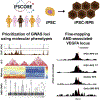
Genomic studies in age-related macular degeneration (AMD) have identified genetic variants that account for the majority of AMD risk. An important next step is to understand the functional consequences and downstream effects of the identified AMD-associated genetic variants. Instrumental for this next step are 'omics' technologies, which enable high-throughput characterization and quantification of biological molecules, and subsequent integration of genomics with these omics datasets, a field referred to as systems genomics. Single cell sequencing studies of the retina and choroid demonstrated that the majority of candidate AMD genes identified through genomic studies are expressed in non-neuronal cells, such as the retinal pigment epithelium (RPE), glia, myeloid and choroidal cells, highlighting that many different retinal and choroidal cell types contribute to the pathogenesis of AMD. Expression quantitative trait locus (eQTL) studies in retinal tissue have identified putative causal genes by demonstrating a genetic overlap between gene regulation and AMD risk. Linking genetic data to complement measurements in the systemic circulation has aided in understanding the effect of AMD-associated genetic variants in the complement system, and supports that protein QTL (pQTL) studies in plasma or serum samples may aid in understanding the effect of genetic variants and pinpointing causal genes in AMD. A recent epigenomic study fine-mapped AMD causal variants by determing regulatory regions in RPE cells differentiated from induced pluripotent stem cells (iPSC-RPE). Another approach that is being employed to pinpoint causal AMD genes is to produce synthetic DNA assemblons representing risk and protective haplotypes, which are then delivered to cellular or animal model systems. Pinpointing causal genes and understanding disease mechanisms is crucial for the next step towards clinical translation. Clinical trials targeting proteins encoded by the AMD-associated genomic loci C3, CFB, CFI, CFH, and ARMS2/HTRA1 are currently ongoing, and a phase III clinical trial for C3 inhibition recently showed a modest reduction of lesion growth in geographic atrophy. The EYERISK consortium recently developed a genetic test for AMD that allows genotyping of common and rare variants in AMD-associated genes. Polygenic risk scores (PRS) were applied to quantify AMD genetic risk, and may aid in predicting AMD progression. In conclusion, genomic studies represent a turning point in our exploration of AMD. The results of those studies now serve as a driving force for several clinical trials. Expanding to omics and systems genomics will further decipher function and causality from the associations that have been reported, and will enable the development of therapies that will lessen the burden of AMD.
Keywords: Age-related macular degeneration; Clinical trial; Complement system; Expression quantitative trait locus; Induced pluripotent stem cells; Omics; Polygenic risk scores; Single cell sequencing; Systems genomics; iPSc-RPE.
Copyright © 2022 The Author(s). Published by Elsevier Ltd.. All rights reserved.
Conflict of interest statement
Declaration of competing interest AIdH is currently an employee of AbbVie; LDO, HHC, HC and LH are employees of Genentech; SK is an employee of Gemini Therapeutics. GSH is a shareholder, consultant, and co-founder of Perceive Biotherapeutics, Inc. and an inventor on patents and patent applications owned by the University of Iowa and the University of Utah. RFM, APV, TS, FG, JLH, JJW, SJT, RA, DS, CCWK, JDB, KAF, BHFW and MBG have no competing interests to declare.
Figures


References
-
- Ajana S, et al., 2021. Predicting progression to advanced age-related macular degeneration from clinical, genetic, and lifestyle factors using machine learning. Ophthalmology 128, 587–597. - PubMed
-
- Biggs R, et al., 2021a. Recombinant complement factor H (GEM103) ocular biodisbribution and activity following intravitreal administration in cynomolgus monkeys. In: 13th International Conference on Complement Therapeutics.
-
- Biggs R, et al., 2020. Age-related macular degeneration associated Complement Factor H variants show impaired functionality in vitro. Invest. Ophthalmol. Vis. Sci. 61, 3699.
-
- Biggs R, et al., 2021b. Mechanism of action of GEM103 (rhCFH) as a therapeutic in AMD. In: 13th International Conference on Complement Therapeutics.
Publication types
MeSH terms
Substances
Grants and funding
LinkOut - more resources
Full Text Sources
Medical
Miscellaneous

