. 2022 Nov:113:1-21.
doi: 10.1016/j.matbio.2022.09.001. Epub 2022 Sep 13.

Human intestinal myofibroblasts deposited collagen VI enhances adhesiveness for T cells - A novel mechanism for maintenance of intestinal inflammation

Affiliations

Human intestinal myofibroblasts deposited collagen VI enhances adhesiveness for T cells - A novel mechanism for maintenance of intestinal inflammation

Si-Nan Lin et al. Matrix Biol. 2022 Nov.

Abstract

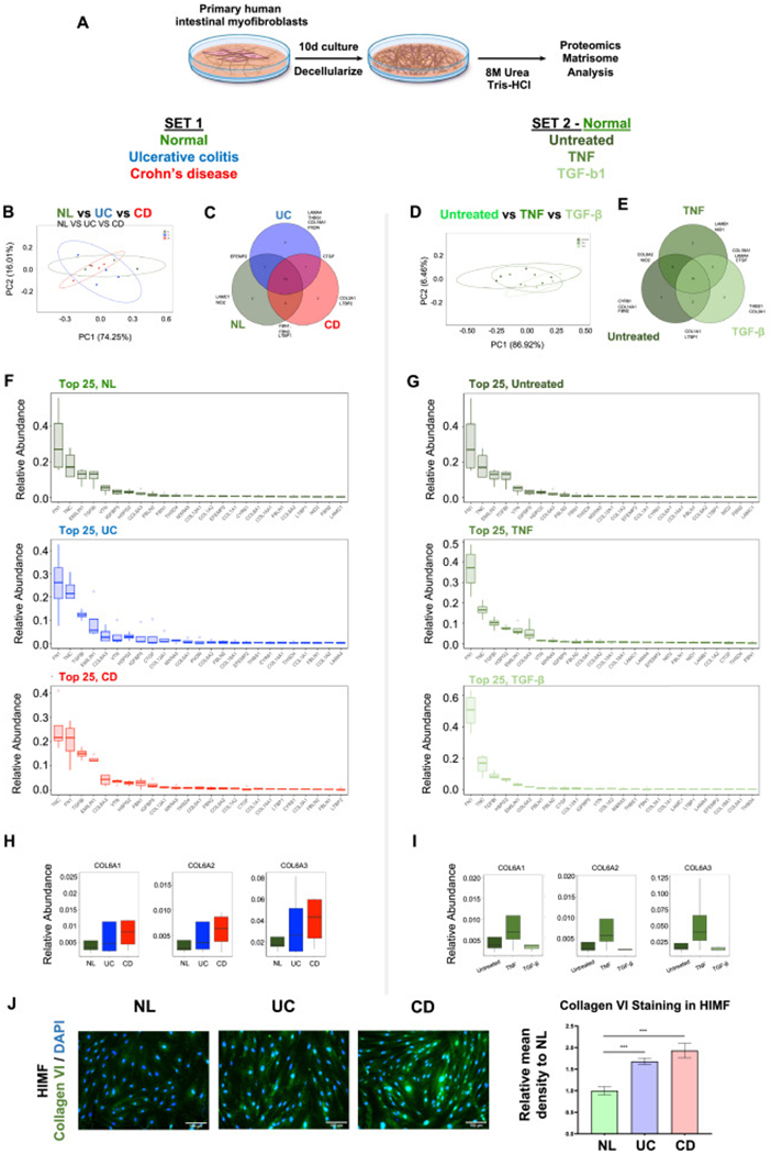
Objective: Inflammatory bowel diseases (IBD) cause chronic intestinal damage and extracellular matrix (ECM) remodeling. The ECM may play an active role in inflammation by modulating immune cell functions, including cell adhesion, but this hypothesis has not been tested in IBD.

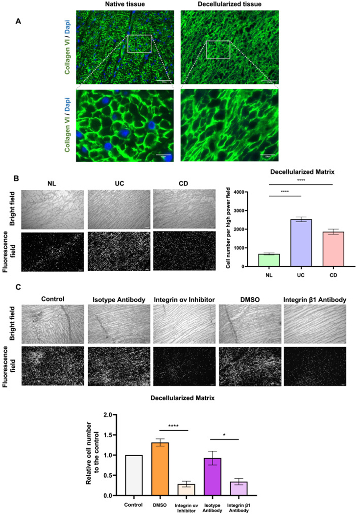
Design: Primary human intestinal myofibroblast (HIMF)-derived ECM from IBD and controls, 3D decellularized colon or ECM molecule-coated scaffolds were tested for their adhesiveness for T cells. Matrisome was analysed via proteomics. Functional integrin blockade was used to investigate the underlying mechanism. Analysis of the pediatric Crohn's disease (CD) RISK inception cohort was used to explore an altered ECM gene expression as a potential predictor for a future complicated disease course.

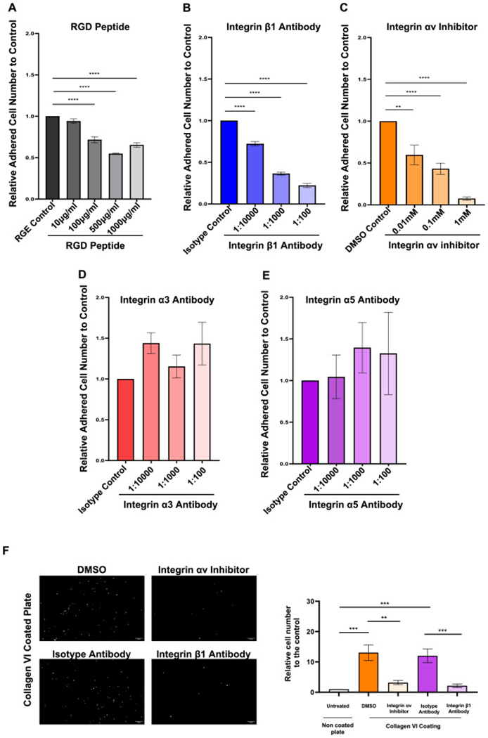
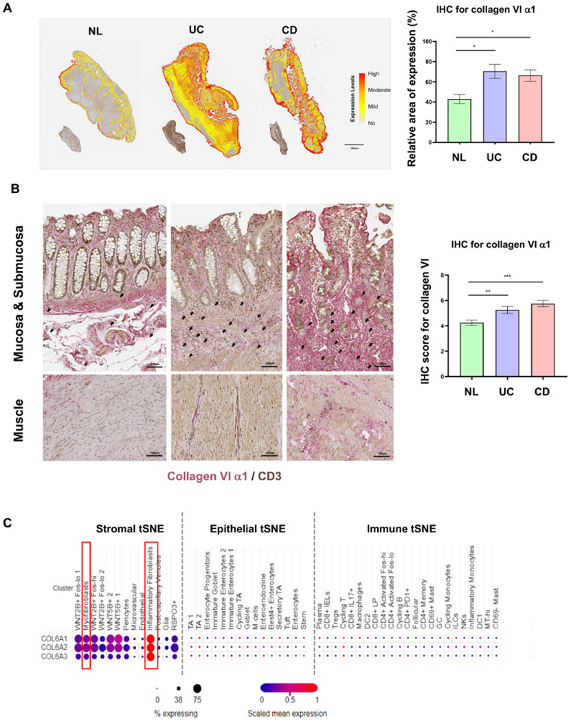
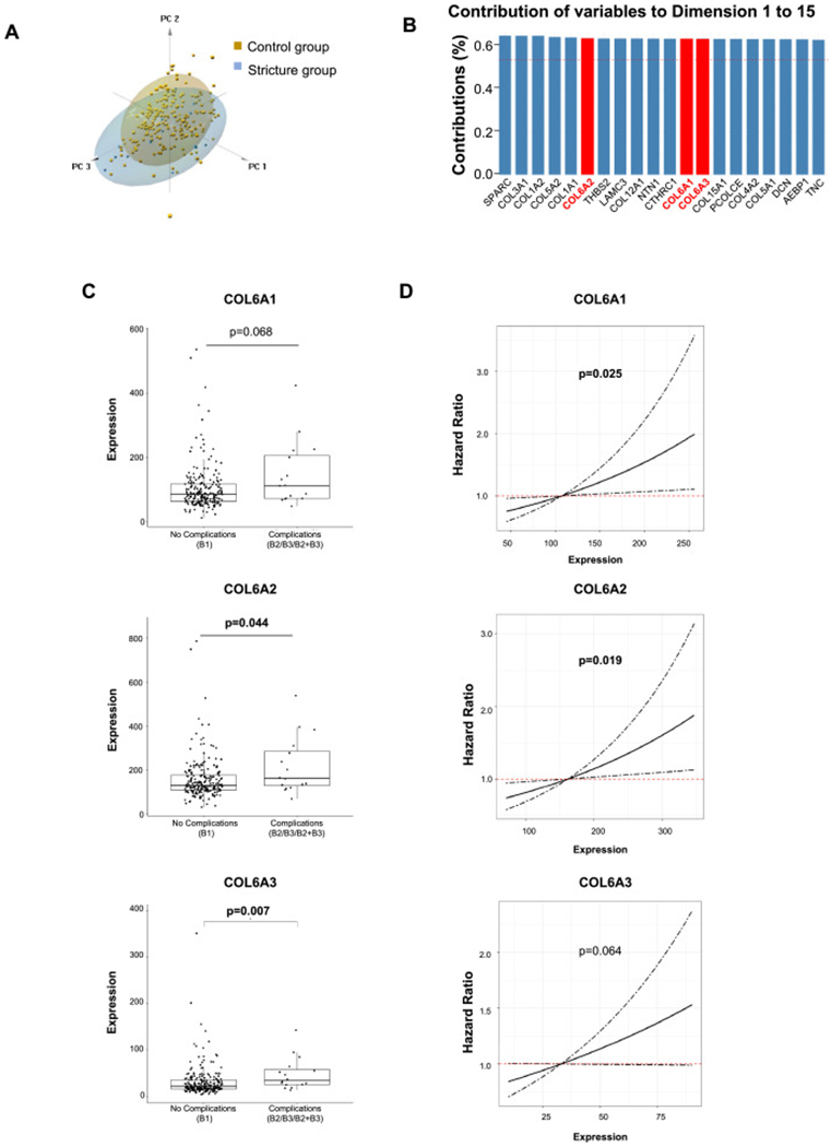
Results: HIMF-derived ECM and 3D decellularized colonic ECM from IBD bound more T cells compared to control. Control HIMFs exposed to the pro-inflammatory cytokines Iinterleukin-1β (IL-1β) and tumor necrosis factor (TNF) increased, and to transforming growth factor-β1 (TGF-β1) decreased ECM adhesiveness to T cells. Matrisome analysis of the HIMF-derived ECM revealed collagen VI as a major culprit for differences in T cell adhesion. Collagen VI knockdown in HIMF reduced adhesion T cell as did the blockage of integrin αvβ1. Elevated gene expression of collagen VI in biopsies of pediatric CD patients was linked to risk for future stricturing disease.

Conclusion: HIMF-derived ECM in IBD binds a remarkably enhanced number of T cells, which is dependent on Collagen VI and integrin αvβ1. Collagen VI expression is a risk factor for a future complicated CD course. Blocking immune cells retention may represent a novel approach to treatment in IBD.

Keywords: Cell adhesion; Extracellular matrix; Inflammatory bowel disease; Intestinal T cells.


Conflict of interest statement

Declaration of Competing Interest F.R. is consultant to Agomab, Allergan, AbbVie, Boehringer-Ingelheim, Celgene, Cowen, Genentech, Gilead, Gossamer, Guidepoint, Helmsley, Index Pharma, Jannsen, Koutif, Metacrine, Morphic, Pfizer, Pliant, Prometheus Biosciences, Receptos, RedX, Roche, Samsung, Takeda, Techlab, Thetis, UCB, 89Bio. C.F. received speaker fees from UCB, Genentech, Sandoz, Janssen and he is consultant for Athos Therapeutics, Inc.

Figures

Fig. 1.
Fig. 1.
Inflammatory bowel disease human intestinal myofibroblast-derived extracellular matrix displays enhanced adhesiveness for T cells. (A) Diagram of the T cell with extracellular matrix (ECM) adhesion assay. (B) An increased number of T cells (MOLT4 on the left, peripheral blood T cells in the middle and lamina propria T cells from a CD patient on the right panel) adhered to inflammatory bowel disease (IBD) human intestinal myofibroblast (HIMF) derived ECM compared to that from non-IBD control (n = 5, t test). (C) No differences in cell number (left) or produced ECM protein amount (right) between HIMF from normal control (NL), ulcerative colitis (UC) and Crohn’s disease (CD) groups were noted during cell culture (n = 5, t test). (D) The adhesiveness of T cells to ECM was increased upon stimulation of NL HIMF with interleukin (IL)-1β and tumor necrosis factor (TNF) but decreased with transforming growth factor (TGF)-β1 in matrix derived from NL HIMF. T cell to ECM adhesion remained unchanged in IBD HIMF exposed to IL-1β and TNF, but was reduced upon HIMF stimulation with interferon (IFN)-γ and TGF-β1 (n = 5, t test). Representative images of T cells adhering to HIMF derived ECM are shown below. *, p < 0.05, **, p < 0.01, ***, p < 0.001, ****, p < 0.0001.
Fig. 2.
Fig. 2.
Matrisome analysis in human intestinal myofibroblast derived matrix reveals distinct extracellular matrix production from control to inflammatory bowel disease, and from untreated to cytokine stimulation. (A) Schematic overview of the process of matrisome analysis. (B) Principal component analysis (PCA) analysis of the human intestinal myofibroblast (HIMF) matrisome showing modest differences among normal control (NL), ulcerative colitis (UC) and Crohn’s disease (CD) groups. (C) Unique extracellular matrix (ECM) components were identified among NL, UC and CD groups. (D) PCA analysis showing minimal differences among untreated, tumor necrosis factor (TNF) stimulated or transforming growth factor (TGF)-β1 exposed NL HIMF in respect to matrisome expression. (E) Unique ECM components were identified among untreated, TNF stimulated, TGF-β1 stimulated NL HIMF. (F) Top 25 abundant matrisome ECM molecules in NL, UC and CD groups. (G) Top 25 abundant matrisome ECM molecules in untreated, TNF stimulated and TGF-β1 stimulated groups. (H) Collagen VI protein expression in UC and CD compared to NL matrisome. (I) Collagen VI protein expression in untreated NL HIMF, or NL HIMF exposed to TNF or TGF-β matrisome. (n = 4 in each group). (J) Immunofluorescence staining of cultured HIMFs showed increased expression of collagen VI α1 in UC and CD compared to NL (n = 6, t test). ***, p < 0.001.
Fig. 3.
Fig. 3.
Collagen VI increases matrix adhesiveness T cells adhesion to extracellular matrix (ECM) coated plates was tested. (A) The adhesiveness of T cells was enhanced with increasing concentrations of collagen VI. Phorbol myristate acetate (PMA) pre-treated T cells showed a higher adherence compared to untreated T cells. (B) The increased adhesiveness of T cells was not only observed in in collagen VI coated plates but also in collagen I and collagen IV coated plates and adhesion increased in all groups after exposure of T cells to PMA (n = 4~6, t test). (C) Human intestinal myofibroblasts (HIMFs) were transfected with small interfering (si)RNA targeting COL6A1 prior to generation of 2D ECM scaffolds. T cell adhesion was robustly decreased in the extracellular matrix (ECM) derived from HIMFs which were transfected by COL6A1 siRNA compared to scrambled siRNA (n = 6, t test). *, p < 0.05, **, p < 0.01, ***, p < 0.001.
Fig. 4.
Fig. 4.
The adhesiveness for T cells is influenced by different cytokines and mediated by integrin αvβ1. T cells adhesion to normal human intestinal myofibroblast (HIMF) derived extracellular matrix (ECM) was tested in the presence or absence of integrin function modulating factors. (A) Arg-Gly-Asp (RGD) inhibited the matrix adhesiveness for T cells in a concentration dependent manner (n = 5, t test). (B)&(C) Blockage of integrin αv and β1 inhibited the adhesiveness in a concentration dependent manner (n = 5, t test). (D)&(E) Blockage of integrin α3 and α5 did not alter the adhesiveness of HIMF derived ECM for T cells (n = 3, t test). (F) The T cell adhesion to collagen VI coated plates was dramatically inhibited by the blockage of integrin αv or integrin β1 (n = 8, t test). **, p < 0.01, ***, p < 0.001, ****, p < 0.0001.
Fig. 5.
Fig. 5.
The expression of collagen VI is elevated in inflamed colon tissue in inflammatory bowel disease. (A) Collagen VI α1 immunohistochemistry of full thickness intestinal tissue sections in ulcerative colitis (UC) and Crohn’s disease (CD), compared to normal control (NL). Automatic quantification by HALO software showed significantly higher expression in UC and CD compared to NL (n = 5, t test). (B) Collagen VI α1 expression on immunohistochemistry was elevated in the mucosa, submucosa and muscle layers in UC and CD, compared to NL. Under high magnification CD3 positive immune cells (brown) were found encased by and in close proximity to collagen VI α1 (red) in the submucosa of inflammatory bowel disease (IBD) tissues (black arrows). The increase in collagen VI α1 in intestinal tissues was confirmed by blinded scoring of full thickness colonic sections using a prespecified scoring system (n = 12, t test). (C) Open access single cell dataset [29] revealed that COL6A1, COL6A2 and COL6A3 were mainly expressed in stromal cells in UC, especially in inflammatory fibroblasts, but not in epithelial and immune cells in UC. *, p < 0.05, **, p < 0.01, ***, p < 0.001.
Fig. 6.
Fig. 6.
T cell adhesiveness of inflammatory bowel disease decellularized tissue is inhibited by the blockage of integrin αv or integrin β1 in decellularized intestinal matrix (A) Immunofluorescence staining showing successful removal of cellular components of the decellularized intestinal tissue as indicated by nuclear DAPI stain with extracellular matrix (ECM) structure and collagen VI amount remaining intact. (B) A higher amount of T cells adhered to ulcerative colitis (UC) and Crohn’s disease (CD) decellularized intestinal tissue sections compared to normal control (NL) tissues (n = 6, t test). (C) The adhesiveness of T cells to decellularized tissue dramatically decreased by the blockage of integrin αv or integrin β1 (n = 6, t test). *, p < 0.05, ****, p < 0.0001.
Fig. 7.
Fig. 7.
Collagen VI gene expression levels are elevated in pediatric Crohn’s disease patients who develop strictures and predicts future stricturing disease. (A) Principal component analysis (PCA) showed modest differences between stricture and control groups. Dimension 1 to 3 were plotted. Scree plot showing the first dimension (component) explained 28.1% variations and the second dimension explained 10.6% variations. (B) COL6A1, COL6A2 and COL6A3 were among the top 20 variables that contribute to the differences between fibrostenotic and control groups from dimension 1 to 15, which explained 80.8% the variations. (C) Both levels of COL6A2 and COL6A3 were higher in the future fibrostenotic group than those in the control group. The level of COL6A1 was nominally higher in fibrostenotic group but did not reach statistical significance. (D) COL6A1 and COL6A2 were risk factors for the future development of strictures. The risk for developing strictures nominally increased with the increase of COL6A3 level but did not reach statistical significance. n=218 in control group and n=16 in stricture group.

References

    1. Guan Q, A comprehensive review and update on the pathogenesis of inflammatory bowel disease, J. Immunol. Res 2019(2019) 7247238. - PMC - PubMed
    1. Rieder F, Fiocchi C, Rogler G, Mechanisms, management, and treatment of fibrosis in patients with inflammatory bowel diseases, Gastroenterology 152 (2) (2017) 340–350 e6. - PMC - PubMed
    1. Mortensen JH, Lindholm M, Langholm LL, Kjeldsen J, Bay-Jensen AC, Karsdal MA, Manon-Jensen T, The intestinal tissue homeostasis - the role of extracellular matrix remodeling in inflammatory bowel disease, Expert Rev. Gastroenterol. Hepatol 13 (10) (2019) 977–993. - PubMed
    1. Shimshoni E, Yablecovitch D, Baram L, Dotan I, Sagi I, ECM remodelling in IBD: innocent bystander or partner in crime? The emerging role of extracellular molecular events in sustaining intestinal inflammation, Gut 64 (3) (2015) 367–372. - PMC - PubMed
    1. Petrey AC, de la Motte CA, The extracellular matrix in IBD: a dynamic mediator of inflammation, Curr. Opin. Gastroenterol 33 (4) (2017) 234–238. - PMC - PubMed

Publication types

Supplementary concepts

LinkOut - more resources