. 2023 Feb;82(2):262-271.
doi: 10.1136/ard-2021-221269. Epub 2022 Sep 15.

Targeting FoxO transcription factors with HDAC inhibitors for the treatment of osteoarthritis

Affiliations

Targeting FoxO transcription factors with HDAC inhibitors for the treatment of osteoarthritis

Hiroki Ohzono et al. Ann Rheum Dis. 2023 Feb.

Abstract

Objectives: Osteoarthritis (OA) features ageing-related defects in cellular homeostasis mechanisms in articular cartilage. These defects are associated with suppression of forkhead box O (FoxO) transcription factors. FoxO1 or FoxO3 deficient mice show early onset OA while FoxO1 protects against oxidative stress in chondrocytes and promotes expression of autophagy genes and the essential joint lubricant proteoglycan 4 (PRG4). The objective of this study was to identify small molecules that can increase FoxO1 expression.

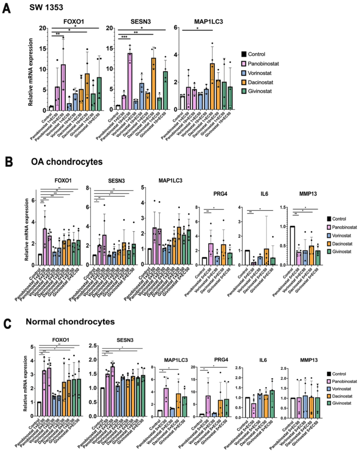
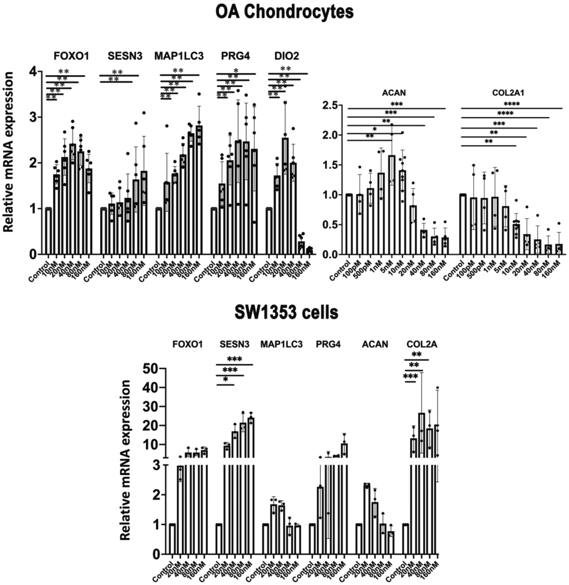
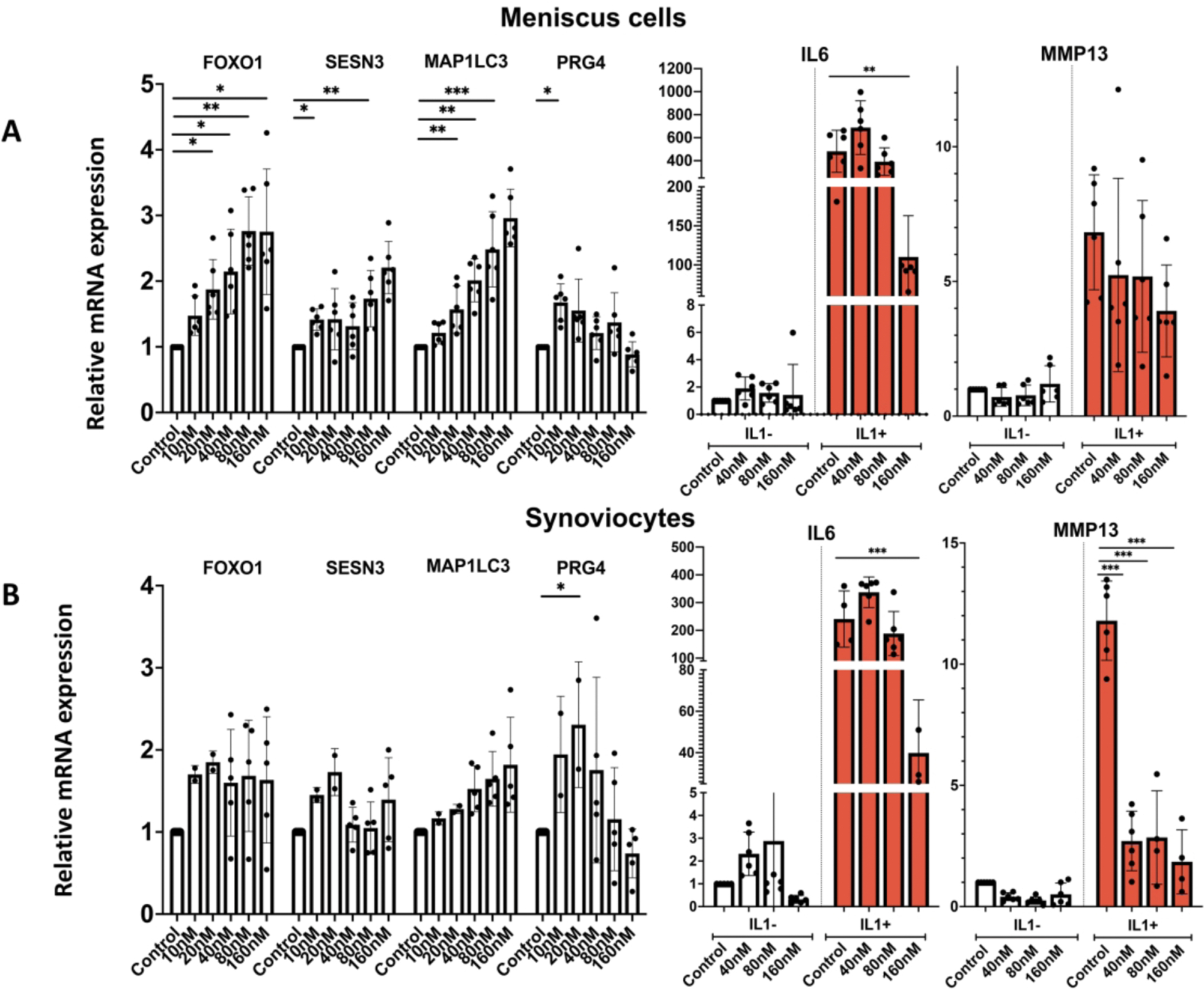
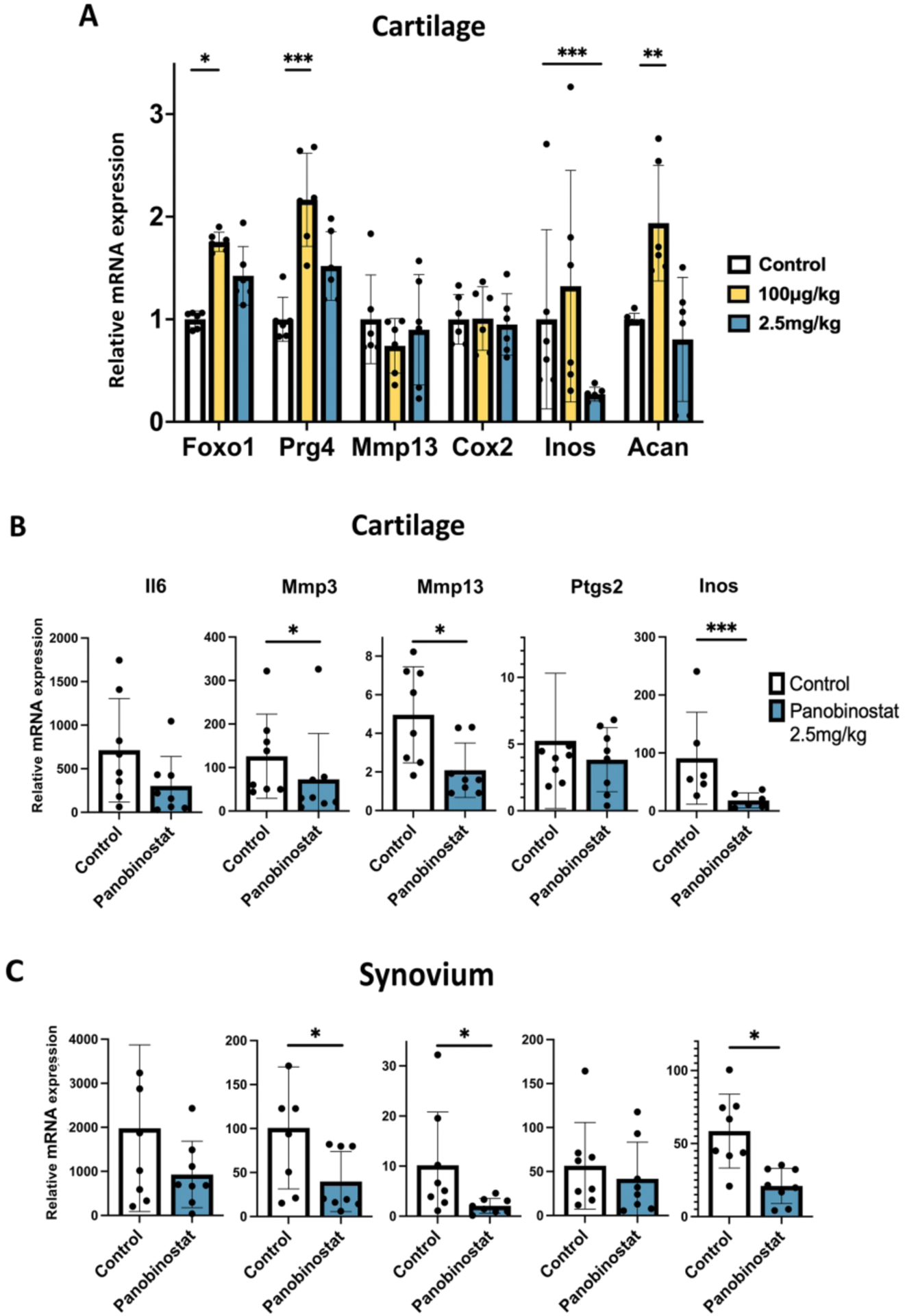
Methods: We constructed a reporter cell line with FoxO1 promoter sequences and performed high-throughput screening (HTS) of the Repurposing, Focused Rescue and Accelerated Medchem (ReFRAME) library . Hits from the HTS were validated and function was assessed in human chondrocytes, meniscus cells and synoviocytes and following administration to mice. The most promising hit, the histone deacetylase inhibitor (HDACI) panobinostat was tested in a murine OA model.

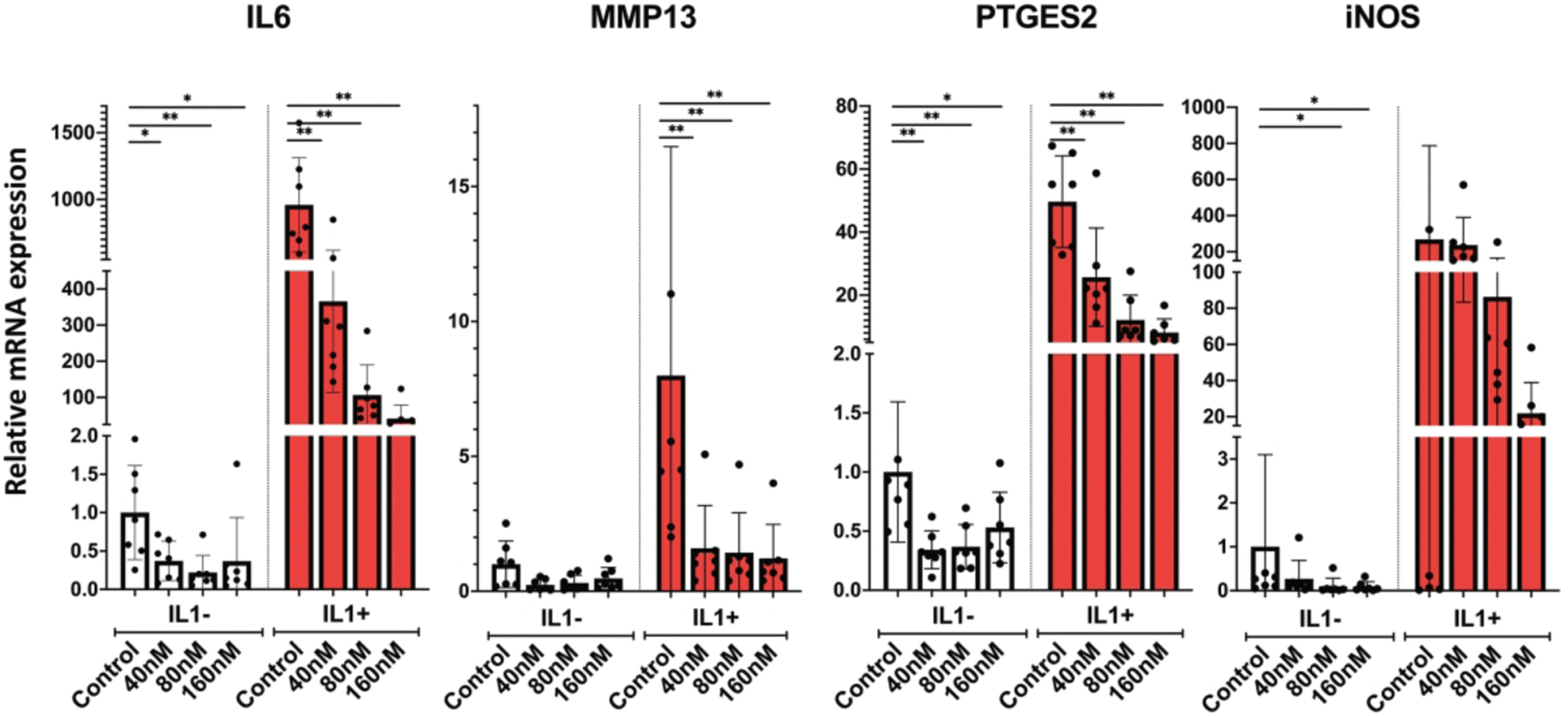
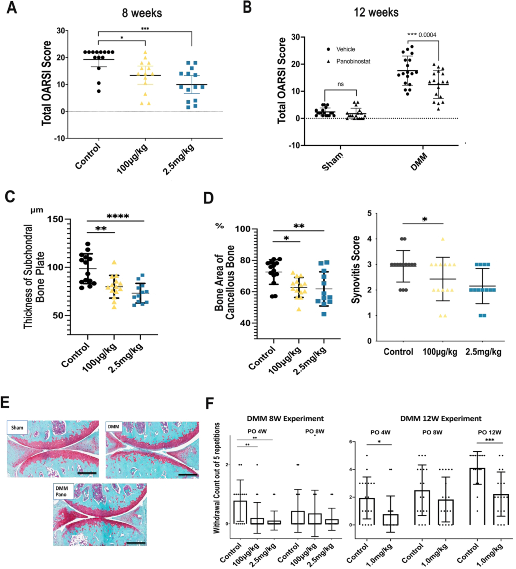
Results: Among the top hits were HDACI and testing in human chondrocytes, meniscus cells and synoviocytes showed that panobinostat was the most promising compound as it increased the expression of autophagy genes and PRG4 while suppressing the basal and IL-1β induced expression of inflammatory mediators and extracellular matrix degrading enzymes. Intraperitoneal administration of panobinostat also suppressed the expression of mediators of OA pathogenesis induced by intra-articular injection of IL-1β. In a murine OA model, panobinostat reduced the severity of histological changes in cartilage, synovium and subchondral bone and improved pain behaviours.

Conclusion: Panobinostat has a clinically relevant activity profile and is a candidate for OA symptom and structure modification.

Keywords: Chondrocytes; Inflammation; Osteoarthritis.


Conflict of interest statement

Competing interests: None declared.

Figures

Figure 1.
Figure 1.. Comparison of top four HDACI.
(A) SW1353 human chondrosarcoma cells were cultured in the presence of the indicated EC50 values of Panobinostat, Vorinostat, Givinostat and Dacinostat for 24 h, and RNA was isolated for qRT-PCR analysis. Independent experiments (N=3) were performed where each condition was tested in duplicate. (B) Human OA chondrocytes (passage 1) from 5 donors were incubated with the indicated doses of Panobinostat, Vorinostat, Givinostat and Dacinostat for 24 hours and RNA was isolated for qRT-PCR analysis. (B) Normal human chondrocytes (passage 1) from 5 donors were incubated with the indicated doses of Panobinostat, Vorinostat, Givinostat and Dacinostat for 24 hours and RNA was isolated for qRT-PCR analysis.
Figure 2.
Figure 2.. Panobinostat and homeostasis and cartilage ECM genes.
(A). Chondrocytes Human OA chondrocytes (passage 1) from 5 donors were incubated with the indicated doses of Panobinostat for 24 h and RNA was isolated for qRT-PCR analysis. (B). SW1353 human chondrosarcoma cells SW1353 cells were treated with the indicated doses of Panobinostat for 24 h and RNA was isolated for qRT-PCR analysis. Independent experiments (N=3) were performed where each condition was tested in duplicate for each condition. ****= p<0.0001; ***= p<0.001; **= p<0.01; *= p<0.05
Figure 3.
Figure 3.. Panobinostat and catabolic genes.
Human OA chondrocytes (passage 1 from 6 donors) were pre-incubated with the indicated doses of Panobinostat for 24 hours. IL-1β (1 ng/ml) was added during the last 6 h and RNA was isolated for qRT-PCR analysis **= p<0.01; *= p<0.05
Figure 4.
Figure 4.. Panobinostat effects on human meniscus cells and synoviocytes.
Human OA meniscus cells at passage 1 from 6 donors (Panel A) and OA synoviocytes at passage 1 from 6 donors (Panel B) were treated with Panobinostat for 24 h when IL-1β (1 ng/ml) was added for additional 6 h and RNA was isolated for RT-qPCR analysis. ***= p<0.001; **= p<0.01; *= p<0.05
Figure 5.
Figure 5.. Panobinostat effects on joint tissues in normal mice.
(A). C57BL/6J mice (n=6) received intraperitoneal injections of Panobinostat on Day 0, day 2 and Day 4 and were euthanized 1–3 h after the last injection. (B). C57BL/6J mice (n=8 each) received one intraperitoneal injection of vehicle and Panobinostat (2.5 mg/kg) and 2 h later IL-1β (50 ng in 10 μl of 5% dextrose) was injected into the right knee and 10 μl of 5% dextrose injected into the left knee. Six h after the IL-1β injection the mice were euthanized. Cartilage and synovium were resected for RNA isolation and RT-qPCR analysis. Data are shown as fold increase in response to IL-1β in Control animals and in Panobinostat treated animals. ***= p<0.001; **= p<0.01; *= p<0.05
Figure 6.
Figure 6.. Analysis of structural changes and symptoms in mice with experimental OA.
Male C57BL/6J mice (n=70), 16-weeks old, were subjected to DMM surgery on the right knee and sham surgery on the left knee. Fourteen mice were randomly assigned to one of three groups, Control (IP injection of 10 μl/g body weight) of 5% dextrose with 0.72% DMSO, Panobinostat 100 μg/kg, and Panobinostat 2.5 mg/kg. IP injections were given every other day starting from one-week post-surgery. Two separate experiments were performed with treatment duration for 8 or 12 weeks after DMM. (A). Knee cartilage changes were scored according to the OARSI system following 8-weeks of Panobinostat treatment (100 μg/kg or 2.5 mg/kg). (B). Knee cartilage changes were scored according to the OARSI system following 12-weeks of Panobinostat treatment (2.5 mg/kg). (C). The area of subchondral bone plate and subchondral cancellous bone were measured by Image J at 8 weeks after DMM. (D). Synovitis score was measured at 8 weeks after DMM. (E). Representative images of Safranin-O stained knee sections from sham surgery control mice, mice with DMM and mice with DMM and Panobinostat (Pano) treatment (2.5 mg/kg). Scale bars, 200 μm (F). Von Frey testing for evaluating mechanical allodynia was performed with 1 g filaments at the indicated time points after DMM in the 8-week and 12-week Panobinostat treatment experiments. ****= p<0.0001; ***= p<0.001; **= p<0.01; *= p<0.05

References

    1. Collins JA, Diekman BO, Loeser RF. Targeting aging for disease modification in osteoarthritis. Curr Opin Rheumatol 2018;30:101–07. - PMC - PubMed
    1. Wang X, Perry TA, Arden N, Chen L, Parsons CM, Cooper C, et al. Occupational Risk in Knee Osteoarthritis: A Systematic Review and Meta-Analysis of Observational Studies. Arthritis Care Res (Hoboken) 2020;72:1213–23. - PMC - PubMed
    1. Courties A, Berenbaum F, Sellam J. The Phenotypic Approach to Osteoarthritis: A Look at Metabolic Syndrome-Associated Osteoarthritis. Joint Bone Spine 2019;86:725–30. - PubMed
    1. Hawker GA. Osteoarthritis is a serious disease. Clin Exp Rheumatol 2019;37 Suppl 120:3–6. - PubMed
    1. Biver E, Berenbaum F, Valdes AM, Araujo de Carvalho I, Bindels LB, Brandi ML, et al. Gut microbiota and osteoarthritis management: An expert consensus of the European society for clinical and economic aspects of osteoporosis, osteoarthritis and musculoskeletal diseases (ESCEO). Ageing Res Rev 2019;55:100946. - PubMed