Validation of a semi-automatic software for optical coherence tomography - analysis in heart transplanted patients
- PMID: 36109445
- PMCID: PMC10519345
- DOI: 10.1007/s10554-022-02722-9
Validation of a semi-automatic software for optical coherence tomography - analysis in heart transplanted patients
Abstract
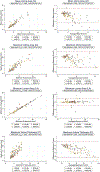
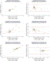
Optical Coherence Tomography (OCT) is an intravascular imaging modality enabling detailed evaluation of cardiac allograft vasculopathy (CAV) after heart transplantation (HTx). However, its clinical application remains hampered by time-consuming manual quantitative analysis. We aimed to validate a semi-automated quantitative OCT analysis software (Iowa Coronary Wall Analyzer, ICWA-OCT) to improve OCT-analysis in HTx patients. 23 patients underwent OCT evaluation of all three major coronary arteries at 3 months (3M) and 12 months (12M) after HTx. We analyzed OCT recordings using the semiautomatic software and compared results with measurements from a validated manual software. For semi-automated analysis, 31,228 frames from 114 vessels were available. The validation was based on a subset of 4287 matched frames. We applied mixed model statistics to accommodate the multilevel data structure with method as a fixed effect. Lumen (minimum, mean, maximum) and media (mean, maximum) metrics showed no significant differences. Mean and maximum intima area were underestimated by the semi-automated method (β-methodmean = - 0.289 mm2, p < 0.01; β-methodmax = - 0.695 mm2, p < 0.01). Bland-Altman analyses showed increasing semi-automatic underestimation of intima measurements with increasing intimal extent. Comparing 3M to 12M progression between methods, mean intimal area showed minor underestimation (β-methodmean = - 1.03 mm2, p = 0.04). Lumen and media metrics showed excellent agreement between the manual and semi-automated method. Intima metrics and progressions from 3M to 12M were slightly underestimated by the semi-automated OCT software with unknown clinical relevance. The semi-automated software has the future potential to provide robust and time-saving evaluation of CAV progression.
Keywords: Cardiac allograft vasculopathy; Heart transplantation; Intravascular imaging; Optical coherence tomography; Validation.
© 2022. The Author(s), under exclusive licence to Springer Nature B.V.
Conflict of interest statement
Figures


References
-
- Clemmensen TS et al. (2013) Twenty years’ experience at the Heart Transplant Center, Aarhus University Hospital, Skejby Denmark. Scand Cardiovasc J 47(6):322–328 - PubMed
-
- Stehlik J et al. (2018) Honoring 50 years of clinical heart transplantation in circulation: in-depth state-of-the-art review. Circulation 137(1):71–87 - PubMed
-
- Clemmensen TS, Jensen NM, Eiskjær H (2021) Imaging of cardiac transplantation: an overview. Semin Nucl Med 51(4):335–348 - PubMed
-
- St Goar FG et al. (1992) Intracoronary ultrasound in cardiac transplant recipients. In vivo evidence of “angiographically silent” intimal thickening. Circulation 85(3):979–987 - PubMed
MeSH terms
Grants and funding
LinkOut - more resources
Full Text Sources
Medical

