. 2023 Feb;48(3):478-488.
doi: 10.1038/s41386-022-01417-y. Epub 2022 Sep 15.

Modulation of methamphetamine memory reconsolidation by neural projection from basolateral amygdala to nucleus accumbens

Affiliations

Modulation of methamphetamine memory reconsolidation by neural projection from basolateral amygdala to nucleus accumbens

Jia-Ying Li et al. Neuropsychopharmacology. 2023 Feb.

Abstract

Drug-associated conditioned cues promote subjects to recall drug reward memory, resulting in drug-seeking and reinstatement. A consolidated memory becomes unstable after recall, such that the amnestic agent can disrupt the memory during the reconsolidation stage, which implicates a potential therapeutic strategy for weakening maladaptive memories. The basolateral amygdala (BLA) involves the association of conditioned cues with reward and aversive valences and projects the information to the nucleus accumbens (NAc) that mediates reward-seeking. However, whether the BLA-NAc projection plays a role in drug-associated memory reactivation and reconsolidation is unknown. We used methamphetamine (MeAM) conditioned place preference (CPP) to investigate the role of BLA-NAc neural projection in the memory reconsolidation. Two weeks before CPP training, we infused adeno-associated virus (AAV) carrying the designer receptor exclusively activated by designer drugs (DREADD) or control constructs. We infused clozapine-N-oxide (CNO) after the recall test to manipulate the neural activity of BLA-NAc projections in mice. We found that after recall, DREADD-mediated inhibition of BLA neurons projecting to the NAc core blunted consolidated MeAM-associated memory. Inhibition of BLA glutamatergic nerve terminals in the NAc core 1 h after recall disrupted consolidated MeAM-associated memory. However, inhibiting this pathway after the time window of reconsolidation failed to affect memory. Furthermore, under the condition without memory retrieval, DREADD-mediated activation of BLA-NAc core projection was required for amnesic agents to disrupt consolidated MeAM-associated memory. Our findings provide evidence that the BLA-NAc pathway activity is involved in the post-retrieval processing of MeAM-associated memory in CPP.


Conflict of interest statement

The authors declare no competing interests.

Figures

Fig. 1
Fig. 1. Anisomycin administration during reconsolidation disrupts MeAM-associated memory.
A Schematic experimental procedure of conditioned place preference (CPP). On Day 1 (baseline), mice were placed into the central chamber and freely explored all three chambers for 15 min. During the training stage (Day 2–6), saline injection paired with the most preferred chamber and then MeAM (2 mg/kg) injection paired with the least preferred chamber 6 h later. After injection, these mice were placed in the respective chamber for 30 min. On Day 7 (the CPP test) and Day 8 (retest), the mice were placed into the central chamber and freely explored all three chambers for 15 min. 1 h after the test on Day 7, the mice were injected with ANI (100 mg/kg) or vehicle (Veh). B The mixed two-way ANOVA showed a significant drug × test interaction effect (F[2,24] = 15.67, p < 0.001), a significant main effect of drug groups (F[1,12] = 12.79, p < 0.01), and a significant main effect of repeated tests (F[2,24] = 24.14, p < 0.001). Bonferroni’s multiple comparison tests showed that all mice preferred to stay in the MeAM-paired chamber (red bars) after the CPP MeAM-paired training (baseline versus CPP test, ***p < 0.001 in vehicle, **p < 0.01 in ANI), indicating that these mice had MeAM-associated memory. Importantly, the retest (green bars) showed that ANI administration after memory retrieval disrupted MeAM-associated memory compared to the vehicle (retest versus CPP test, ***p < 0.001 in ANI-treated mice, p > 0.05 in vehicle-treated mice). n = 7 per group. C The experimental timeline of ANI infusion into the NAc core. Another group of mice was infused ANI (1 µg in 0.8 µL/side) or the vehicle into the NAc core 30 min after the CPP test. n = 7 and 8 in the vehicle and ANI group, respectively. D The mixed two-way ANOVA showed a significant drug × test interaction effect (F[2,26] = 13.47, p < 0.001), a significant main effect of drug groups (F[1,13] = 11.32, p < 0.01), and a significant main effect of repeated tests (F[2,26] = 43.58, p < 0.001). Post-hoc Bonferroni’s tests showed that ANI infusion into the NAc core after memory retrieval disrupted MeAM-associated memory compared to the vehicle (retest versus CPP test, ***p < 0.001 in ANI-infused mice, p > 0.05 in vehicle-infused mice). MeAM methamphetamine, CPP conditioned place preference, Veh vehicle, ANI anisomycin. Data represent mean ± SEM.
Fig. 2
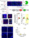
Fig. 2. Post-retrieval chemogenetic inhibition of the BLA neurons projecting to the NAc disrupts the consolidated MeAM-associated memory.
A (Top) schematic representation showed that the retrograde tracer, red retrobeads, infused into the NAc core. (Bottom) Quantification of images showed that retrobead signals were present in the BLA (40 ± 1.83%), the prefrontal cortex (PFC, 31 ± 1.53%), and the ventral tegmental area-substantia nigra (VTA-SN, 17.1 ± 1.11%). Minor signals were also observed in CA1 of the ventral hippocampus (8.88 ± 0.8%), the basomedial amygdala (BMA), and the ventromedial hypothalamus (VMH). The one-way ANOVA indicated that the BLA was the major input of the NAc core (F(4, 103) = 146.9, p < 0.001; BLA versus PFC, p < 0.001 in Bonferroni’s multiple comparisons test). Brain slices from three mice. Data represent mean ± SEM. B Representative images showed the injection site of retrobeads in the NAc core. Scale bar represents 550 µm. The distribution of retrobeads included the BLA, the PFC, the VTA, the CA1, the BMA, and the VMH. The CPu was a negative control brain area with no retrobead signals. Scale bar represents 200 µm. NAc, the nucleus accumbens; BLA, the basolateral amygdala; PFC, the prefrontal cortex; VTA, the ventral tegmental area; CA1, the ventral hippocampal CA1; others include the basomedial amygdala (BMA) and the ventromedial hypothalamus (VMH). CPu, the caudate putamen. C Schematic representation of the virus infusion and cannula implantation sites and the experimental procedure. CNO (2 µg/side) or vehicle was infused 1 h after the test on Day 7. The mice were sacrificed for histology analysis 90 min after the retest. D Representative images showed the retrograde AAV infusion site in the NAc core (left) and the reporter mCherry in the BLA (right). Scale bars represent 550 µm and 100 µm in the NAc core and in the BLA, respectively. E In the CPP procedure, all mice after CPP training preferred the MeAM-paired chamber (baseline versus CPP test, p < 0.05 and p < 0.001 in control and hM4Di-expressing mice, respectively, Bonferroni’s multiple comparisons test). In the retest on Day 8 (green bars), intra-BLA CNO infusion reduced the preference for the MeAM-paired chamber compared to the vehicle infusion in hM4Di-expressing mice (retest versus CPP test, p < 0.001 in CNO infusion, Bonferroni’s multiple comparisons test). CNO infusion into the BLA had no effect in transduced control viral mice (retest versus CPP test, p > 0.05, Bonferroni’s multiple comparisons test). The mixed two-way ANOVA in hM4Di-expressing mice, the drug × test interaction, F[2,28] = 15.65, p < 0.001, n = 7 and 9 in vehicle and CNO, respectively; the repeated one-way ANOVA in control mice, F(1.8, 9.2) = 18.62, p < 0.001, n = 6. F Representative images showed c-Fos expression on hM4Di-expressing BLA neurons in the vehicle- and CNO-infused mice. The scale bar represents 40 µm. Green, c-Fos; red, mCherry; blue, DAPI. G CNO infusion decreased retest-induced c-Fos expression on hM4Di-expressing neurons in the BLA compared to the vehicle. Vehicle versus CNO, p < 0.001, unpaired t test. n = 7 and 9 mice in vehicle and CNO, respectively. H CNO infusion also decreased retest-induced c-Fos-positive cells in the NAc core compared to the vehicle. The bottom images show the magnification of the white dotted box in the top images. Vehicle versus CNO, p < 0.001, unpaired t-test. n = 7 and 9 mice in vehicle and CNO, respectively. The scale bars represent 200 and 100 µm in the top and bottom images, respectively. Green, c-Fos; blue, DAPI. Data represent mean ± SEM.
Fig. 3
Fig. 3. Chemogenetic inhibition of nerve terminals of glutamatergic BLA neurons in the NAc core after retrieval disrupts the consolidated MeAM-associated memory.
A The retrograde virus was infused in the NAc core, and the mCherry reporter expression was analyzed in the BLA. B Representative images showed that mCherry signals (red) colocalized with CaMKIIα signals (green), the glutamatergic neuron marker, in the BLA. Triangles pointed to the colocalized cells. Scale bar represents 40 µm. The insets on the right: amplification images for the mCherry-only positive cell (top), the colocalized cell (middle), and the CaMKII-only positive cell (bottom). C Image quantification revealed that 76.48% of mCherry-positive BLA neurons were CaMKIIα-positive. n = 8 brain slices from three mice. D Schematic representation of the virus infusion and cannula implantation sites and the experimental procedure. CNO (2 µg/side) or vehicle was infused into the NAc core 1 h after the CPP test. E Representative images showed the anterograde AAV infusion site in the BLA (left) and its amplification image (right). Scale bar represents 550 µm (left) and 200 µm (right). F In hM4Di-expressing mice, CNO infusion reduced the preference for the MeAM-paired chamber in the retest compared to vehicle infusion, suggesting that inhibiting the nerve terminals of glutamatergic BLA neurons in the NAc core during reconsolidation disrupted MeAM-associated memory. Mixed two-way ANOVA, the drug × test interaction, F[2,22] = 6.88, p < 0.01; n = 5 and 8 in vehicle and CNO, respectively; retest versus test, p < 0.01 in CNO, Bonferroni’s multiple comparisons test. By contrast, in control EGFP-expressing mice, CNO infusion had no effect on the retest. Repeated one-way ANOVA, F(1.8, 7.2) = 21.59, p < 0.01; n = 5; retest versus test, p > 0.05, Bonferroni’s multiple comparisons test. G Confocal images showed fiber-like mCherry-labeled BLA axon terminals in contact with c-Fos-positive cells in the NAc core. The scale bar represents 50 µm in the left and 10 µm in the right magnification photos. H Representative images showed c-Fos-positive cells (green) in the NAc core (top) and in the shell (bottom) of the vehicle and CNO-infused mice. The scale bar represents 100 µm. I Image quantification revealed that CNO infusion reduced c-Fos expression in the NAc core compared to vehicle infusion. CNO infusion had no effects on the NAc shell. CNO versus vehicle in the NAc core, p < 0.001, unpaired t-test, n = 14 brain slices from four mice; the NAc shell, p > 0.05, n = 7 and 8 brain slices from four mice. Data represent mean ± SEM.
Fig. 4
Fig. 4. Delayed inhibition of BLA neuronal nerve terminals in the NAc core after the time window of memory reconsolidation fails to affect the consolidated MeAM-associated memory.
A Diagram of the virus infusion and cannulas implantation sites and the experimental procedure. CNO (2 µg/side) or vehicle was infused into the NAc core 18 h after the CPP test. B Delayed CNO infusion failed to affect MeAM-associated memory in hM4Di-expressing mice. Mixed two-way ANOVA, the drug ×  test interaction, F[2,18] = 0.05, p = 0.95; n = 5 and 6 in vehicle and CNO, respectively; retest versus test, p > 0.05, Bonferroni’s multiple comparisons test. Data represent mean ± SEM.
Fig. 5
Fig. 5. Activating the glutamatergic BLA-NAc core pathway is sufficient for anisomycin to disrupt MeAM-associated memory.
A Schematic representation of the virus infusion and cannula implantation sites and the experimental procedure. Twenty-four hours after the CPP test, the mice received CNO infusion into the NAc core (Day 8) to activate nerve terminals of glutamatergic BLA neurons. 1 h later, ANI was infused into the NAc core, and the mice did not experience a memory test. On the next day (Day 9), the mice received the memory retest. B Representative images showed the anterograde AAV infusion site in the BLA (left) and its amplification image (right). Scale bar represents 550 µm (left) and 200 µm (right). C ANI-only or CNO-only infusion on Day 8 failed to affect MeAM-associated memory in the retest. The combination of CNO infusion with ANI infusion disrupted the consolidated MeAM-associated memory of hM3Dq-expressing mice in the retest, suggesting that ANI disrupted the MeAM-associated memory only when activating the BLA-NAc core pathway. Mixed two-way ANOVA, the drug × test interaction, F [4,36] = 12.57, p < 0.001; n = 7 per group; retest versus CPP test, p > 0.05 in Veh+ANI and CNO + veh, p < 0.001 in CNO + ANI, Bonferroni’s multiple comparisons test. D, E Several days after the CPP experiments, these hM3Dq mice received CNO infusion into the NAc core and then were sacrificed for histology 90 min later. CNO infusion induced c-Fos expression on mCherry-positive neurons in the BLA of hM3Dq-expressing mice compared to vehicle infusion. CNO versus vehicle, p < 0.05, unpaired t-test, n = 6 brain slices from three mice per group. F Representative images showed c-Fos-positive cells (green) in the NAc core. CNO infusion induced c-Fos expression in the NAc core compared to the vehicle. The bottom images show the magnification of the white dotted box in the top images. Vehicle versus CNO, p < 0.001, unpaired t-test. n = 10 brain slices from three mice per group. The scale bars represent 200 and 100 µm in the top and bottom images, respectively. Green, c-Fos; blue, DAPI. Data represent mean ± SEM.

Similar articles

Cited by

References

    1. Everitt BJ, Robbins TW. Neural systems of reinforcement for drug addiction: from actions to habits to compulsion. Nat Neurosci. 2005;8:1481–9. doi: 10.1038/nn1579. - DOI - PubMed
    1. Gipson CD, Kupchik YM, Shen H, Reissner KJ, Thomas CA, Kalivas PW. Relapse induced by cues predicting cocaine depends on rapid, transient synaptic potentiation. Neuron. 2013;77:867–72. doi: 10.1016/j.neuron.2013.01.005. - DOI - PMC - PubMed
    1. Sampedro-Piquero P, Ladrón de Guevara-Miranda D, Pavón FJ, Serrano A, Suárez J, Rodríguez de Fonseca F, et al. Neuroplastic and cognitive impairment in substance use disorders: a therapeutic potential of cognitive stimulation. Neurosci Biobehav Rev. 2019;106:23–48. doi: 10.1016/j.neubiorev.2018.11.015. - DOI - PubMed
    1. Bossert JM, Liu SY, Lu L, Shaham Y. A role of ventral tegmental area glutamate in contextual cue-induced relapse to heroin seeking. J Neurosci. 2004;24:10726–30. doi: 10.1523/jneurosci.3207-04.2004. - DOI - PMC - PubMed
    1. Bossert JM, Poles GC, Wihbey KA, Koya E, Shaham Y. Differential effects of blockade of dopamine D1-family receptors in nucleus accumbens core or shell on reinstatement of heroin seeking induced by contextual and discrete cues. J Neurosci. 2007;27:12655–63. doi: 10.1523/jneurosci.3926-07.2007. - DOI - PMC - PubMed

Publication types

Substances