Review
. 2023 Mar;20(3):145-167.
doi: 10.1038/s41569-022-00759-w. Epub 2022 Sep 15.

Inflammatory signalling in atrial cardiomyocytes: a novel unifying principle in atrial fibrillation pathophysiology

Affiliations
Review

Inflammatory signalling in atrial cardiomyocytes: a novel unifying principle in atrial fibrillation pathophysiology

Dobromir Dobrev et al. Nat Rev Cardiol. 2023 Mar.

Abstract

Inflammation has been implicated in atrial fibrillation (AF), a very common and clinically significant cardiac rhythm disturbance, but its precise role remains poorly understood. Work performed over the past 5 years suggests that atrial cardiomyocytes have inflammatory signalling machinery - in particular, components of the NLRP3 (NACHT-, LRR- and pyrin domain-containing 3) inflammasome - that is activated in animal models and patients with AF. Furthermore, work in animal models suggests that NLRP3 inflammasome activation in atrial cardiomyocytes might be a sufficient and necessary condition for AF occurrence. In this Review, we evaluate the evidence for the role and pathophysiological significance of cardiomyocyte NLRP3 signalling in AF. We first summarize the evidence for a role of inflammation in AF and review the biochemical properties of the NLRP3 inflammasome, as defined primarily in studies of classic inflammation. We then briefly consider the broader evidence for a role of inflammatory signalling in heart disease, particularly conditions that predispose individuals to develop AF. We provide a detailed discussion of the available information about atrial cardiomyocyte NLRP3 inflammasome signalling in AF and related conditions and evaluate the possibility that similar signalling might be important in non-myocyte cardiac cells. We then review the evidence on the role of active resolution of inflammation and its potential importance in suppressing AF-related inflammatory signalling. Finally, we consider the therapeutic potential and broader implications of this new knowledge and highlight crucial questions to be addressed in future research.


Conflict of interest statement

The authors declare no competing interests.

Figures

Fig. 1
Fig. 1. Pathways involved in NLRP3 inflammasome activation.
Molecular pathways involved in NLRP3 inflammasome activation on the basis of information from classic models of inflammation. NLRP3 inflammasome activation in response to tissue stress and damage involves inflammasome priming (Signal 1), leading to increased expression of the major inflammasome components (NLRP3, ASC and caspase 1), and triggering (Signal 2), promoting assembly of the NLRP3 inflammasome. Subsequent activation of caspase 1 leads to activation of several proteins including IL-1β and IL-18, as well as gasdermin D (GSDMD) and IL-1β release through pores formed by the GSDMD amino terminus (NT) fragment. ATR, angiotensin receptor; BRCC36, Lys-63-specific deubiquitinase 36; CaMKII, Ca2+/calmodulin-dependent protein kinase II; CARD, caspase recruitment domain; CaSR, calcium-sensing receptor; DAMP, damage-associated molecular pattern; ER, endoplasmic reticulum; FADD, FAS-associated death domain protein; GSDMD-CT, gasdermin D carboxy terminus; IL-1R, IL-1 receptor type 1; IP3, inositol 1,4,5-trisphosphate; IP3R, inositol 1,4,5-trisphosphate receptor; JNK, JUN N-terminal kinase 1; LPS, lipopolysaccharide; LRR, leucine-rich repeat; mtDNA, mitochondrial DNA; MyD88, myeloid differentiation primary response protein 88; NEK7, NIMA related kinase 7; NF-κB, nuclear factor-κB; NLRP3, NACHT-, LRR- and pyrin domain-containing 3; NOD, nucleotide-binding oligomerization domain-containing protein; Ox-mtDNA, oxidized mitochondrial DNA; P2X7, P2X purinoceptor 7; PAMP, pathogen-associated molecular pattern; PAR4, proteinase-activated receptor 4; PIP2, phosphatidylinositol 4,5-bisphosphate; PKA, protein kinase A; PLC, phospholipase C; PP2A, protein phosphatase 2A; PYD, pyrin domain; ROS, reactive oxygen species; RIPK1, receptor-interacting serine/threonine-protein kinase 1; RYR2, ryanodine receptor 2; SR, sarcoplasmic reticulum; TLR, Toll-like receptor; TNF, tumour necrosis factor; TNFR, tumour necrosis factor receptor; TRP, transient receptor potential; Ub, ubiquitin.
Fig. 2
Fig. 2. Pathways leading to NLRP3 inflammasome activation induced by comorbidities and cardiovascular risk factors.
Main pathways through which risk factors, heart diseases and comorbidities can promote NLRP3 inflammasome activation, potentially leading to atrial fibrillation and contributing to myocardial pathology in conditions such as acute myocardial infarction and heart failure. Risk factors, including gut microbiota dysbiosis, obesity, diabetes mellitus, right heart disease and coronary artery disease, promote the production of several mediators that activate transmembrane receptors involved in NLRP3 inflammasome priming, including Toll-like receptor 4 (TLR4), tumour necrosis factor receptors (TNFRs) and IL-1 receptor type 1 (IL-1R1), as well as downstream activation of signalling pathways dependent on Ca2+/calmodulin-dependent protein kinase II (CaMKII), reactive oxygen species (ROS) and nuclear factor-κB (NF-κB). Assembly of inflammasome components (triggering) is mediated, among other pathways, by P2X purinoceptor 7 (P2X7) signalling and K+ depletion, as well as histone deacetylase 6 (HDAC6). Downstream activation of caspase 1 and formation of pores by the gasdermin D (GSDMD) amino terminus (NT) fragment subsequently allows the release of IL-1β and IL-18 from the cell. In parallel, these pathways promote pro-arrhythmic atrial remodelling, including spontaneous Ca2+ release events, delayed afterdepolarizations and triggered activity, as well as re-entry-promoting shortening of action potential duration and effective refractory period in combination with structural remodelling and associated conduction abnormalities. Dashed arrows indicate indirect effects. DAMP, damage-associated molecular pattern; GSDMD-CT, gasdermin D carboxy terminus; LPS, lipopolysaccharide; NLRP3, NACHT-, LRR- and pyrin domain-containing 3.
Fig. 3
Fig. 3. Inflammation resolution concept.
In response to injury or infection, acute inflammation is activated to favour the return to homeostasis. During the initiation phase, damage signals (pathogen-associated molecular patterns (PAMPs) and damage-associated molecular patterns (DAMPs) promote phospholipase A2 (PLA2)-induced accumulation of arachidonic acid (AA), activation of the nuclear factor-κB (NF-κB) pathway and assembly of the NLRP3 inflammasome, leading to the release of pro-inflammatory substances, recruitment of polymorphonuclear leukocytes (PMNs) and monocyte polarization towards pro-inflammatory M1 macrophages. Apoptotic PMNs and M1 macrophages secrete 12-lipoxygenase (12-LOX) and 15-LOX, which triggers lipid-mediator class switching, signalled by increased production of resolution-promoting mediators from AA, eicosapentaenoic acid (EPA) and docosahexaenoic acid (DHA). Pro-resolution autacoids orchestrate the inhibition of PMN infiltration, the increased release of anti-inflammatory cytokines (IL-10 and IL-13) and increased macrophage polarization to anti-inflammatory M2 macrophages to phagocytize damaged cells. Inflammation resolution is characterized by efferocytosis, tissue repair, wound healing, preservation of functions and homeostasis. Failed resolution of inflammation is associated with dysfunctional triggering of lipid-mediator class switching, perpetuation of production and secretion of pro-inflammatory mediators (IL-1β, IL-6 and IL-18), aggravation and chronicity of the inflammatory status, development of fibrosis, myocardial damage, loss of myocardial function and arrhythmogenesis. CaMKII, calcium/calmodulin-dependent kinase II; COX, cyclooxygenase; CYP450, cytochrome P450; LTB4, leukotriene B4, LXA4, lipoxin A4; MAPK, mitogen-activated protein kinase; NLRP3, NACHT-, LRR- and pyrin domain-containing 3; PGE2, prostaglandin E2; PGI2, prostaglandin I2; ROS, reactive oxygen species; TXA2, thromboxane A2.
Fig. 4
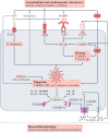
Fig. 4. Cardiomyocyte–immune cell interactions promote and maintain atrial fibrillation.
Dynamic interactions between cardiac cells and immune cells create a pro-inflammatory substrate for the promotion and maintenance of atrial fibrillation. Atrial fibrillation-promoting risk factors activate various mediators that signal through pattern-recognition receptors (PRRs) on cardiomyocytes to activate the cardiomyocyte NLRP3 inflammasome, leading to the release of IL-1β and IL-18 and activation of signalling pathways dependent on Ca2+/calmodulin-dependent protein kinase II (CaMKII), reactive oxygen species (ROS) and nuclear factor-κB (NF-κB). Subsequent activation of IL-1 receptor type 1 (IL-1R1) on macrophages promotes feedforward amplification of inflammatory signalling, whereas IL-1R1-mediated stimulation of fibroblasts causes extracellular matrix remodelling. Together, these processes result in cardiomyocyte Ca2+ handling abnormalities, including ryanodine receptor 2 (RYR2) channel remodelling, delayed afterdepolarizations, and triggered action potentials and ectopic activity, as well as re-entry-promoting effective refractory period (ERP) shortening and fibrotic structural remodelling, thereby inducing the principal mechanisms responsible for atrial fibrillation. AngII, angiotensin II; ANP, atrial natriuretic peptide; DAMPs, damage-associated molecular patterns; LPS, lipopolysaccharide; GSDMD, gasdermin D; GSDMD-NT, gasdermin D amino terminus; NLRP3, NACHT-, LRR- and pyrin domain-containing 3; Thr, thrombin; TNF, tumour necrosis factor.

References

    1. Andrade J, Khairy P, Dobrev D, Nattel S. The clinical profile and pathophysiology of atrial fibrillation: relationships among clinical features, epidemiology, and mechanisms. Circ. Res. 2014;114:1453–1468. doi: 10.1161/CIRCRESAHA.114.303211. - DOI - PubMed
    1. Heijman J, Guichard JB, Dobrev D, Nattel S. Translational challenges in atrial fibrillation. Circ. Res. 2018;122:752–773. doi: 10.1161/CIRCRESAHA.117.311081. - DOI - PubMed
    1. Chung MK, et al. C-reactive protein elevation in patients with atrial arrhythmias: inflammatory mechanisms and persistence of atrial fibrillation. Circulation. 2001;104:2886–2891. doi: 10.1161/hc4901.101760. - DOI - PubMed
    1. Harada M, Van Wagoner DR, Nattel S. Role of inflammation in atrial fibrillation pathophysiology and management. Circ. J. 2015;79:495–502. doi: 10.1253/circj.CJ-15-0138. - DOI - PMC - PubMed
    1. Gong T, Liu L, Jiang W, Zhou R. DAMP-sensing receptors in sterile inflammation and inflammatory diseases. Nat. Rev. Immunol. 2020;20:95–112. doi: 10.1038/s41577-019-0215-7. - DOI - PubMed

Publication types

Substances