

Oligo-Fucoidan supplementation enhances the effect of Olaparib on preventing metastasis and recurrence of triple-negative breast cancer in mice
- PMID: 36109724
- PMCID: PMC9479298
- DOI: 10.1186/s12929-022-00855-6
Oligo-Fucoidan supplementation enhances the effect of Olaparib on preventing metastasis and recurrence of triple-negative breast cancer in mice
Abstract
Background: Seaweed polysaccharides have been recommended as anticancer supplements and for boosting human health; however, their benefits in the treatment of triple-negative breast cancers (TNBCs) and improving immune surveillance remain unclear. Olaparib is a first-in-class poly (ADP-ribose) polymerase inhibitor. Oligo-Fucoidan, a low-molecular-weight sulfated polysaccharide purified from brown seaweed (Laminaria japonica), exhibits significant bioactivities that may aid in disease management.
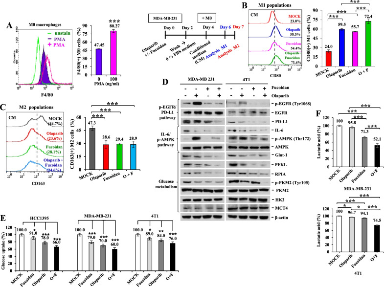
Methods: Macrophage polarity, clonogenic assays, cancer stemness properties, cancer cell trajectory, glucose metabolism, the TNBC 4T1 cells and a 4T1 syngeneic mouse model were used to inspect the therapeutic effects of olaparib and Oligo-Fucoidan supplementation on TNBC aggressiveness and microenvironment.
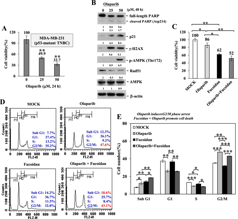
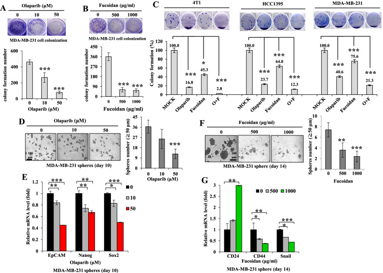
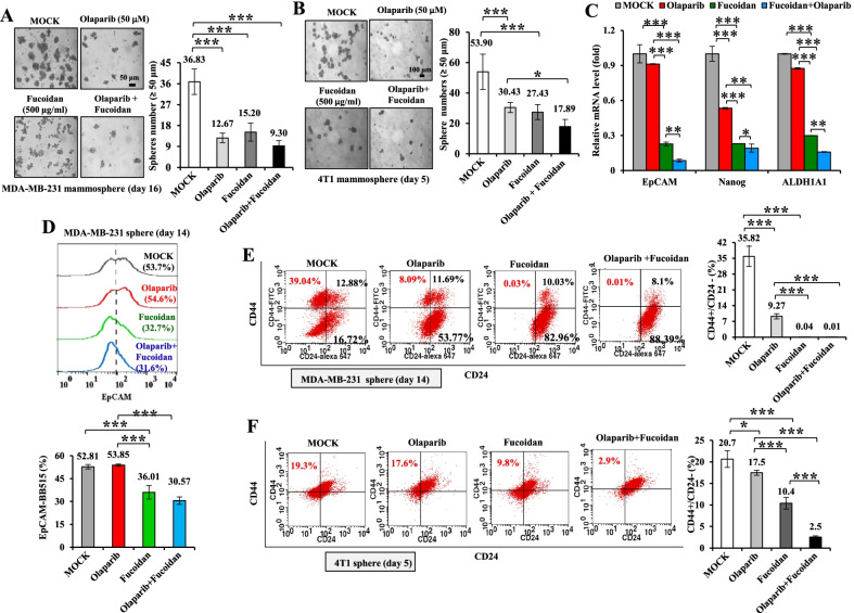
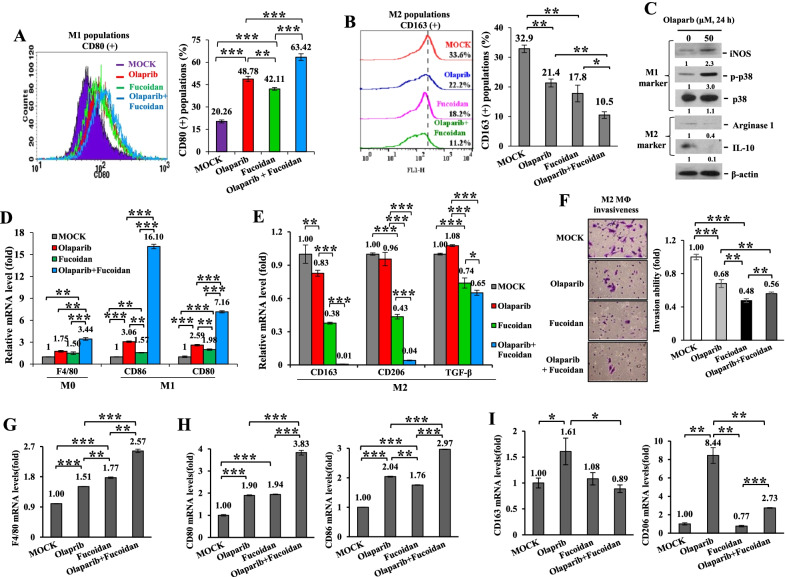
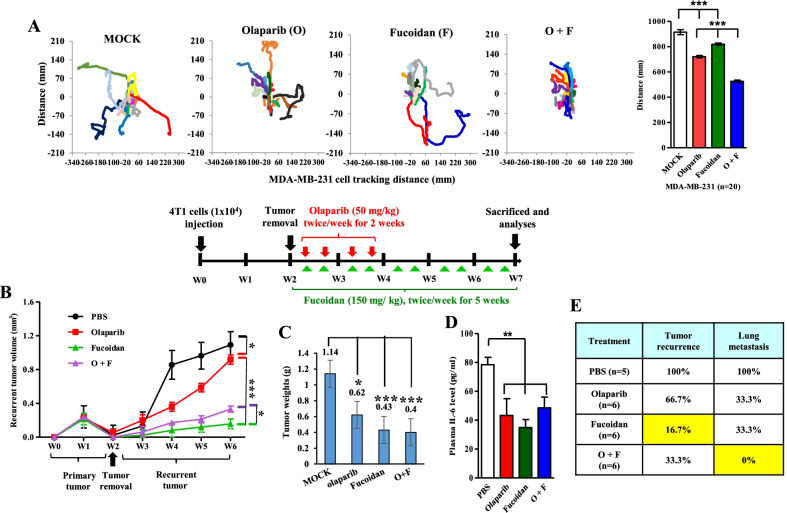
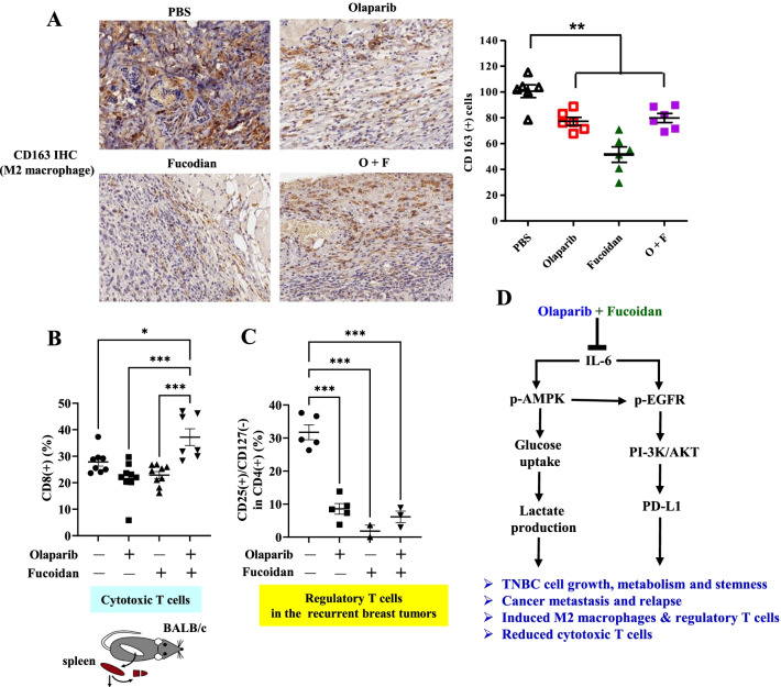
Results: Olaparib treatment increased sub-G1 cell death and G2/M arrest in TNBC cells, and these effects were enhanced when Oligo-Fucoidan was added to treat the TNBC cells. The levels of Rad51 and programmed death-ligand 1 (PD-L1) and the activation of epidermal growth factor receptor (EGFR) and adenosine 5'-monophosphate (AMP)-activated protein kinase (AMPK) facilitate drug resistance and TNBC metastasis. However, the combination of olaparib and Oligo-Fucoidan synergistically reduced Rad51 and PD-L1 levels, as well as the activity of EGFR and AMPK; consistently, TNBC cytotoxicity and stemness were inhibited. Oligo-Fucoidan plus olaparib better inhibited the formation of TNBC stem cell mammospheroids with decreased subpopulations of CD44high/CD24low and EpCAMhigh cells than monotherapy. Importantly, Oligo-Fucoidan plus olaparib repressed the oncogenic interleukin-6 (IL-6)/p-EGFR/PD-L1 pathway, glucose uptake and lactate production. Oligo-Fucoidan induced immunoactive and antitumoral M1 macrophages and attenuated the side effects of olaparib, such as the promotion on immunosuppressive and protumoral M2 macrophages. Furthermore, olaparib plus Oligo-Fucoidan dramatically suppressed M2 macrophage invasiveness and repolarized M2 to the M0-like (F4/80high) and M1-like (CD80high and CD86high) phenotypes. In addition, olaparib- and Oligo-Fucoidan-pretreated TNBC cells resulted in the polarization of M0 macrophages into CD80(+) M1 but not CD163(+) M2 macrophages. Importantly, olaparib supplemented with oral administration of Oligo-Fucoidan in mice inhibited postsurgical TNBC recurrence and metastasis with increased cytotoxic T cells in the lymphatic system and decreased regulatory T cells and M2 macrophages in tumors.
Conclusion: Olaparib supplemented with natural compound Oligo-Fucoidan is a novel therapeutic strategy for reprogramming cancer stemness, metabolism and the microenvironment to prevent local postsurgical recurrence and distant metastasis. The combination therapy may advance therapeutic efficacy that prevent metastasis, chemoresistance and mortality in TNBC patients.
Keywords: Cancer stem cells; Glucose uptake; IL-6/EGFR/PD-L1 signaling pathway; Lactate production; M1/M2 macrophage polarization; Olaparib; Oligo-Fucoidan; Triple-negative breast cancer.
© 2022. The Author(s).
Conflict of interest statement
The authors declare no competing interests.
Figures
Similar articles
-
Histone deacetylase inhibitor, suberoylanilide hydroxamic acid (SAHA), enhances anti-tumor effects of the poly (ADP-ribose) polymerase (PARP) inhibitor olaparib in triple-negative breast cancer cells.Breast Cancer Res. 2015 Mar 7;17:33. doi: 10.1186/s13058-015-0534-y. Breast Cancer Res. 2015. PMID: 25888415 Free PMC article.
-
Immunotherapeutic IL-6R and targeting the MCT-1/IL-6/CXCL7/PD-L1 circuit prevent relapse and metastasis of triple-negative breast cancer.Theranostics. 2024 Mar 3;14(5):2167-2189. doi: 10.7150/thno.92922. eCollection 2024. Theranostics. 2024. PMID: 38505617 Free PMC article.
-
Taraxacum mongolicum extract inhibited malignant phenotype of triple-negative breast cancer cells in tumor-associated macrophages microenvironment through suppressing IL-10 / STAT3 / PD-L1 signaling pathways.J Ethnopharmacol. 2021 Jun 28;274:113978. doi: 10.1016/j.jep.2021.113978. Epub 2021 Mar 11. J Ethnopharmacol. 2021. PMID: 33716082
-
Triple negative breast cancer: Key role of Tumor-Associated Macrophages in regulating the activity of anti-PD-1/PD-L1 agents.Biochim Biophys Acta Rev Cancer. 2018 Jan;1869(1):78-84. doi: 10.1016/j.bbcan.2017.10.007. Epub 2017 Nov 7. Biochim Biophys Acta Rev Cancer. 2018. PMID: 29126881 Review.
-
State of art of advanced triple negative breast cancer.Breast J. 2019 Sep;25(5):967-970. doi: 10.1111/tbj.13369. Epub 2019 Jun 2. Breast J. 2019. PMID: 31155832 Review.
Cited by
-
Therapeutic Efficacy, Radiotoxicity and Abscopal Effect of BNCT at the RA-3 Nuclear Reactor Employing Oligo-Fucoidan and Glutamine as Adjuvants in an Ectopic Colon Cancer Model in Rats.Life (Basel). 2023 Jul 11;13(7):1538. doi: 10.3390/life13071538. Life (Basel). 2023. PMID: 37511913 Free PMC article.
-
Fucoidan-decorated metal-zoledronic acid nanocomplexes suppress tumor metastasis by inducing ferroptotic cell death and enhancing cancer immunotherapy.J Nanobiotechnology. 2025 Jun 2;23(1):405. doi: 10.1186/s12951-025-03473-0. J Nanobiotechnology. 2025. PMID: 40452005 Free PMC article.
-
Upregulation of p300 in paclitaxel-resistant TNBC: implications for cell proliferation via the PCK1/AMPK axis.Pharmacogenomics J. 2024 Feb 20;24(2):5. doi: 10.1038/s41397-024-00324-3. Pharmacogenomics J. 2024. PMID: 38378770
-
NOS2 and COX2 Provide Key Spatial Targets that Determine Outcome in ER- Breast Cancer.bioRxiv [Preprint]. 2023 Dec 23:2023.12.21.572859. doi: 10.1101/2023.12.21.572859. bioRxiv. 2023. PMID: 38187532 Free PMC article. Preprint.
-
Manganese and IL-12 treatment alters the ovarian tumor microenvironment.Aging (Albany NY). 2024 Jan 3;16(1):191-206. doi: 10.18632/aging.205361. Epub 2024 Jan 3. Aging (Albany NY). 2024. PMID: 38175694 Free PMC article.
References
-
- Oliveras-Ferraros C, Vazquez-Martin A, Lopez-Bonet E, Martin-Castillo B, Del Barco S, Brunet J, et al. Growth and molecular interactions of the anti-EGFR antibody cetuximab and the DNA cross-linking agent cisplatin in gefitinib-resistant MDA-MB-468 cells: new prospects in the treatment of triple-negative/basal-like breast cancer. Int J Oncol. 2008;33(6):1165–1176. - PubMed
MeSH terms
Substances
Grants and funding
LinkOut - more resources
Full Text Sources
Research Materials
Miscellaneous
