. 2022 Sep 15;20(1):138.
doi: 10.1186/s12958-022-00998-z.

Circ-CSNK1G1 promotes cell proliferation, migration, invasion and glycolysis metabolism during triple-negative breast cancer progression by modulating the miR-28-5p/LDHA pathway

Affiliations

Circ-CSNK1G1 promotes cell proliferation, migration, invasion and glycolysis metabolism during triple-negative breast cancer progression by modulating the miR-28-5p/LDHA pathway

Xiaochen Zan et al. Reprod Biol Endocrinol. .

Abstract

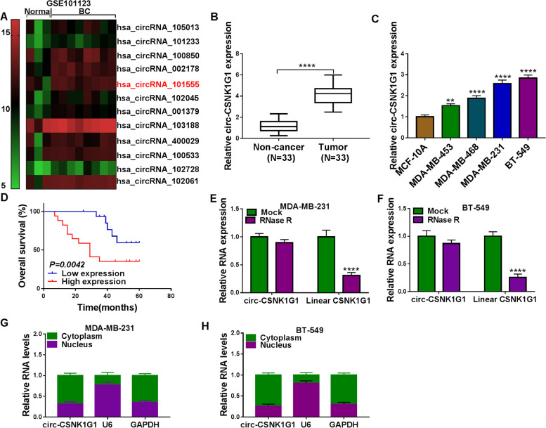
Background: Circular RNAs (circRNAs) play a vital role in cancer progression. However, there are still numerous circRNAs that have not been functionally explored. Our study aimed to disclose the role of circ-CSNK1G1 in triple-negative breast cancer (TNBC).

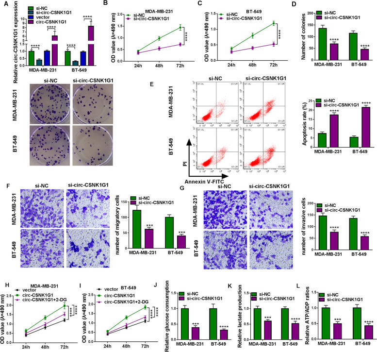
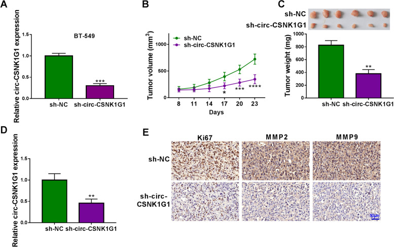
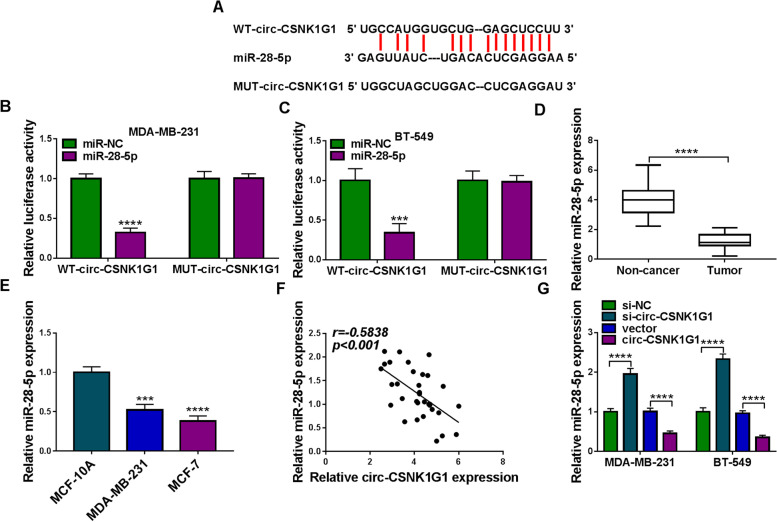
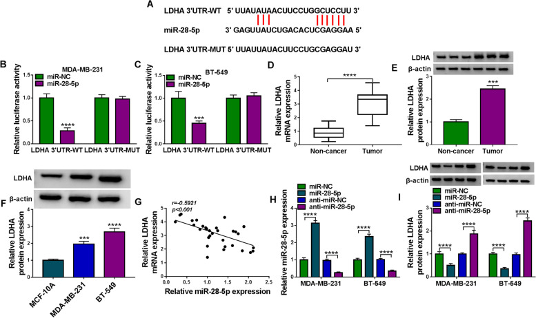
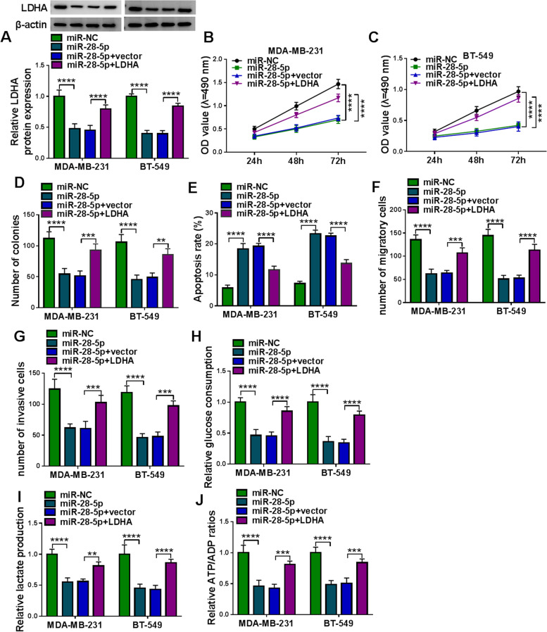
Methods: The expression of circ-CSNK1G1, miR-28-5p and lactate dehydrogenase A (LDHA) mRNA was measured by quantitative real-time polymerase chain reaction (qPCR), and the expression of LDHA protein was measured by western blot. Cell proliferation was assessed using MTT assay and colony formation assay. Cell apoptosis was monitored using flow cytometry assay. Cell migration and cell invasion were investigated using transwell assay. Glycolysis progression was assessed according to glucose consumption, lactate production and ATP/ADP ratio. Tumor formation assay in nude mice was conducted to verify the role of circ-CSNK1G1 in vivo. The interplays between miR-28-5p and circ-CSNK1G1 or LDHA were confirmed by dual-luciferase reporter assay.

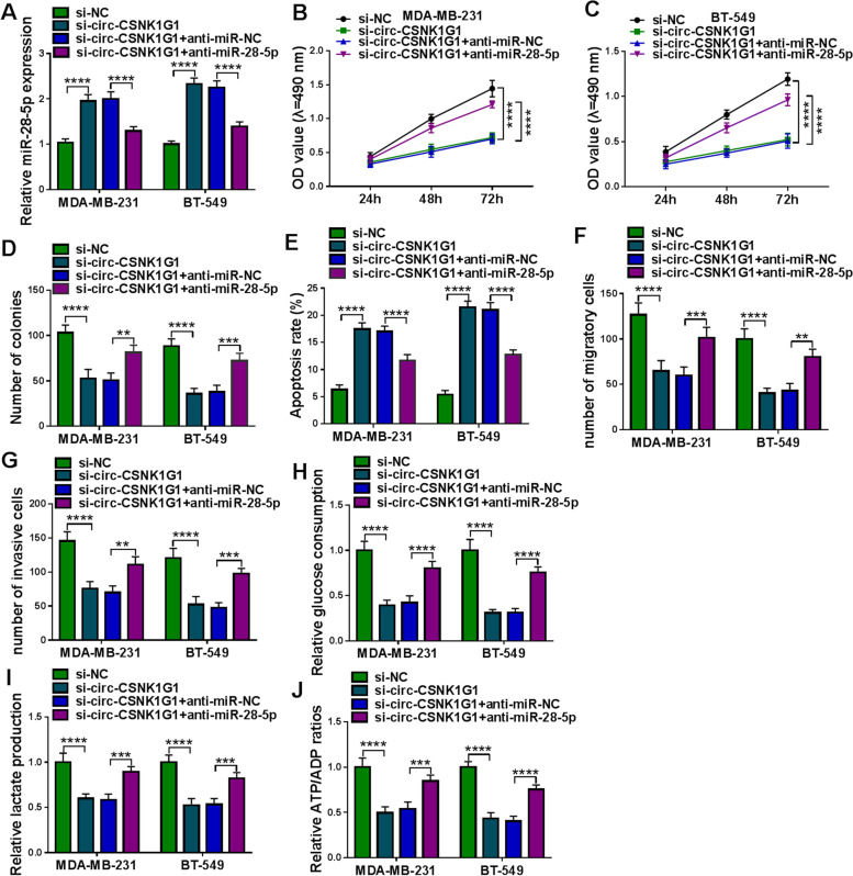
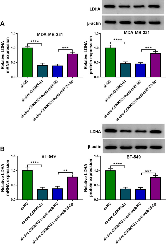
Results: Circ-CSNK1G1 was upregulated in TNBC tissues and cells. Circ-CSNK1G1 knockdown suppressed cancer cell proliferation, migration, invasion and glycolysis energy metabolism, promoted cell apoptosis in vitro, and blocked tumor growth in vivo. Mechanism analysis showed that circ-CSNK1G1 positively regulated LDHA expression by suppressing miR-28-5p. Rescue experiments presented that circ-CSNK1G1 played functions by targeting miR-28-5p, and miR-28-5p participated in TNBC progression by degrading LDHA.

Conclusion: Circ-CSNK1G1 promotes cell proliferation, migration, invasion and glycolysis metabolism during TNBC development by regulating the miR-28-5p/LDHA pathway.

Keywords: LDHA; Triple-negative breast cancer; circ-CSNK1G1; miR-28-5p.


Conflict of interest statement

The authors declare that they have no competing interests.

Figures

Fig. 1
Fig. 1
Circ-CSNK1G1 was upregulated in TNBC tissues and cells. A The data of circRNA expression in TNBC tissues were collected from a circRNA expression profile (GEO accession: GSE101123). B The expression of circ-CSNK1G1 in tumor tissues and normal breast tissues was detected by qPCR. C The expression of circ-CSNK1G1 in TNBC cell lines and non-cancer cell line was detected by qPCR. D Overall survival was analyzed by Kaplan-Meier plot and log-rank test. E and F The stability of circ-CSNK1G1 was ensured using RNase R test. G and H The location of circ-CSNK1G1 was analyzed by qPCR. **P < 0.01 and ****P < 0.0001
Fig. 2
Fig. 2
Circ-CSNK1G1 silence inhibited cell proliferation, migration, invasion and glycolysis. A The efficiency of circ-CSNK1G1 silence and overexpression was examined by qPCR. B and C MTT assay was conducted to assess proliferation in MDA-MB-231 and BT-549 cells after circ-CSNK1G1 silence. D Colony formation assay was used to assess proliferation in cells after circ-CSNK1G1 silence. E Flow cytometry assay was performed to monitor cell apoptosis in cells after circ-CSNK1G1 silence. F and G Transwell assay was performed to monitor cell migration and invasion in cells after circ-CSNK1G1 silence. H and I The effects of 2-DG on the role of circ-CSNK1G1 were determined by MTT assay. J-L Glucose consumption, lactate production and ATP/ADP ratios were detected to assess glycolysis progression. ***P < 0.001 and ****P < 0.0001
Fig. 3
Fig. 3
Circ-CSNK1G1 knockdown blocked tumor growth. A The expression of circ-CSNK1G1 in BT-549 cells after sh-circ-CSNK1G1 or sh-NC transfection was measured by qPCR. B and C Tumor volume and tumor weight were measured to monitor tumor growth. D The expression of circ-CSNK1G1 in the removed tumor tissues was measured by qPCR. (E) The abundance of Ki67, MMP2 and MMP9 in tumor tissues was analyzed by IHC assay. **P < 0.01 and ***P < 0.001
Fig. 4
Fig. 4
Circ-CSNK1G1 suppressed the expression of miR-28-5p. A The binding sites between circ-CSNK1G1 and miR-28-5p. B and C Luciferase reporter assay was performed to confirm the relationship between circ-CSNK1G1 and miR-28-5p. D The expression of miR-28-5p in tumor tissues and normal tissues was detected by qPCR. E The expression of miR-28-5p in MCF-10 A, MDA-MB-231 and BT-549 cells was detected by qPCR. F The correlation between circ-CSNK1G1 and miR-28-5p expression was analyzed by Pearson analysis. G The effects of circ-CSNK1G1 silence and overexpression on miR-28-5p expression were detected by qPCR. ***P < 0.001 and ****P < 0.0001
Fig. 5
Fig. 5
MiR-28-5p deficiency recovered breast cell malignant behaviors suppressed by circ-CSNK1G1 silence. Rescue experiments were performed in MDA-MB-231 and BT-549 cells transfected with si-circ-CSNK1G1 alone or si-circ-CSNK1G1 + anti-miR-28-5p together, using si-NC or si-circ-CSNK1G1 + anti-miR-NC as the control. A The expression of miR-28-5p was detected using qPCR. B-D Cell proliferation was analyzed using MTT assay and colony formation assay. E Cell apoptosis was assessed using flow cytometry assay. F and G Cell migration and cell invasion were investigated using transwell assay. H-J Glucose consumption, lactate production and ATP/ADP ratios were detected to assess glycolysis progression. **P < 0.01, ***P < 0.001, and ****P < 0.0001
Fig. 6
Fig. 6
MiR-28-5p suppressed the expression of LDHA. A The binding sites between miR-28-5p and LDHA 3’UTR. B and C Dual-luciferase reporter assay was performed to confirm the interaction between miR-28-5p and LDHA. D and E The expression of LDHA mRNA and protein in tumor tissues and normal tissues was measured by qPCR and western blot. F The expression of LDHA protein in MCF-10 A, MDA-MB-231 and BT-549 cells was measured by western blot. G The correlation between LDHA mRNA expression and miR-28-5p expression was analyzed by Pearson analysis. H and I The effects of miR-28-5p overexpression and deficiency on the expression of LDHA mRNA and protein were determined by qPCR and western blot. ***P < 0.001 and ****P < 0.0001
Fig. 7
Fig. 7
LDHA overexpression recovered TNBC cell malignant behaviors suppressed by miR-28-5p restoration. Rescue experiments were performed in MDA-MB-231 and BT-549 cells transfected with miR-28-5p alone or miR-28-5p + LDHA together, using miR-NC or miR-28-5p + vector as the control. A The expression of LDHA protein in these transfected cells was detected by western blot. B-D Cell proliferation was assessed using MTT assay and colony formation assay. E Cell apoptosis was monitored using flow cytometry assay. (F and G) Cell migration and cell invasion were determined using transwell assay. H-J Glycolysis metabolism progression was assessed by detecting glucose consumption, lactate production and ATP/ADP ratios. **P < 0.01, ***P < 0.001, and ****P < 0.0001
Fig. 8
Fig. 8
Circ-CSNK1G1 decoyed miR-28-5p to release LDHA. A and B The effects of circ-CSNK1G1 knockdown alone or circ-CSNK1G1 knockdown combined with miR-28-5p inhibition on the expression of LDHA was detected using qPCR and western blot. **P < 0.01, ***P < 0.001, and ****P < 0.0001

References

    1. Siegel RL, Miller KD, Jemal A. Cancer statistics, 2019. CA Cancer J Clin. 2019;69(1):7–34. doi: 10.3322/caac.21551. - DOI - PubMed
    1. Pasculli B, Barbano R, Parrella P. Epigenetics of breast cancer: Biology and clinical implication in the era of precision medicine. Semin Cancer Biol. 2018;51:22–35. doi: 10.1016/j.semcancer.2018.01.007. - DOI - PubMed
    1. Mathe A, et al. Novel genes associated with lymph node metastasis in triple negative breast cancer. Sci Rep. 2015;5:15832. doi: 10.1038/srep15832. - DOI - PMC - PubMed
    1. Nicolini A, Ferrari P, Duffy MJ. Prognostic and predictive biomarkers in breast cancer: Past, present and future. Semin Cancer Biol. 2018;52(Pt 1):56–73. doi: 10.1016/j.semcancer.2017.08.010. - DOI - PubMed
    1. Wang X, Fang L. Advances in circular RNAs and their roles in breast Cancer. J Exp Clin Cancer Res. 2018;37(1):206. doi: 10.1186/s13046-018-0870-8. - DOI - PMC - PubMed

LinkOut - more resources