Review
. 2022 Aug;74(4):433-457.
doi: 10.1007/s10616-022-00539-x. Epub 2022 Jul 24.

Status in molluscan cell line development in last one decade (2010-2020): impediments and way forward

Affiliations
Review

Status in molluscan cell line development in last one decade (2010-2020): impediments and way forward

Soumya Balakrishnan et al. Cytotechnology. 2022 Aug.

Abstract

Despite the attempts that have started since the 1960s, not even a single cell line of marine molluscs is available. Considering the vast contribution of marine bivalve aquaculture to the world economy, the prevailing viral threats, and the dismaying lack of advancements in molluscan virology, the requirement of a marine molluscan cell line is indispensable. This synthetic review discusses the obstacles in developing a marine molluscan cell line concerning the choice of species, the selection of tissue and decontamination, and cell culture media, with emphasis given on the current decade 2010-2020. Detailed accounts on the experiments on the virus cultivation in vitro and molluscan cell immortalization, with a brief note on the history and applications of the molluscan cell culture, are elucidated to give a holistic picture of the current status and future trends in molluscan cell line development.

Supplementary information: The online version contains supplementary material available at 10.1007/s10616-022-00539-x.

Keywords: Cell line; Culture medium; Decontamination; Immortalization; Molluscan cell culture; Transfection; Transformation; Virus.


Conflict of interest statement

Competing interestThe authors declare that they have no competing interests.

Figures

Fig. 1
Fig. 1
Marine molluscan groups used for cell culture development (In % of the selected publications 2010–2020)
Fig. 2
Fig. 2
Tissues used for marine molluscan cell culture development and type of study. M mantle, L larva, DG digestive gland, HC hemocytes, H heart, GD gonad, G gills, N neuron, LP labial palps, R radula, AM adductor muscle (results from 60 experiments)
Fig. 3
Fig. 3
Experiments indicating number of subcultures performed for each type of tissue. X-axis (numbers 0, 1, 2, 3, 4, 5) represent the number of subcultures (results from 60 experiments)
Fig. 4
Fig. 4
Experiments indicating longevity of cell culture derived from each type of tissue. M mantle, L larva, DG digestive gland, HC hemocytes, H heart, GD gonad, G gills, N neuron, LP labial palps, R radula, AM adductor muscle (results from 60 experiments)
Fig. 5
Fig. 5
Depuration of animals (with or without antibiotics) and sterilization of tissue for the initiation of marine molluscan cell culture. M mantle, L larva, DG digestive gland, HC hemocytes, H heart, GD gonad, G gills, N neuron, LP labial palps, R radula, AM adductor muscle, Depurated with Ab- depuration of animals in the presence of antibiotics, Ab wash- tissue washed with antibiotics (results from 60 experiments)
Fig. 6
Fig. 6
Cell culture medium used for the development of marine molluscan cell culture. L-15-Leibovitz's L-15 Medium, SW-seawater, HBSS-Hank's Balanced Salt Solution, Opti-MEM-Opti-MEM® I reduced serum media (results from 60 experiments)
Fig. 7
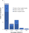
Fig. 7
FBS/FCS (in %) used as a supplement in cell culture medium for the development of marine molluscan cell culture (results from 60 experiments)

References

    1. Alavi MR, Fernández-Robledo JA, Vasta GR. Development of an in vitro assay to examine intracellular survival of Perkinsus marinus trophozoites upon phagocytosis by oyster (Crassostrea virginica and Crassostrea ariakensis) hemocytes. J Parasitol. 2009;95:900–907. doi: 10.1645/GE-1864.1. - DOI - PubMed
    1. Arzul I, Corbeil S, Morga B, Renault T. Viruses infecting marine molluscs. J Invertebr Pathol. 2017;147:118–135. doi: 10.1016/j.jip.2017.01.009. - DOI - PubMed
    1. Awaji M, Machii A. Fundamental studies on in vivo and in vitro pearl formation—contribution of outer epithelial cells of pearl oyster mantle and pearl sacs. Aqua-BioSci Monogr. 2011;4:1–39. doi: 10.5047/absm.2011.00401.0001. - DOI
    1. Balbi T, Ciacci C, Grasselli E, et al. Utilization of Mytilus digestive gland cells for the in vitro screening of potential metabolic disruptors in aquatic invertebrates. Comp Biochem Physiol Part C Toxicol Pharmacol. 2017;191:26–35. doi: 10.1016/j.cbpc.2016.08.009. - DOI - PubMed
    1. Barrick A, Guillet C, Mouneyrac C, Châtel A. Investigating the establishment of primary cultures of hemocytes from Mytilus edulis. Cytotechnology. 2018;70:1205–1220. doi: 10.1007/s10616-018-0212-x. - DOI - PMC - PubMed

LinkOut - more resources