

The mechanism of action of a novel neuroprotective low molecular weight dextran sulphate: New platform therapy for neurodegenerative diseases like Amyotrophic Lateral Sclerosis
- PMID: 36110516
- PMCID: PMC9468270
- DOI: 10.3389/fphar.2022.983853
The mechanism of action of a novel neuroprotective low molecular weight dextran sulphate: New platform therapy for neurodegenerative diseases like Amyotrophic Lateral Sclerosis
Abstract
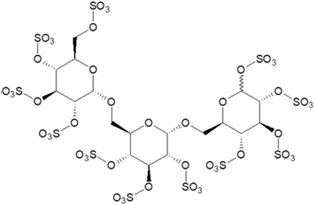
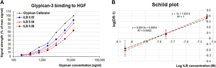
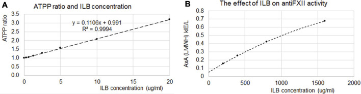
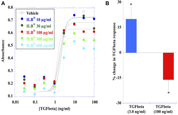
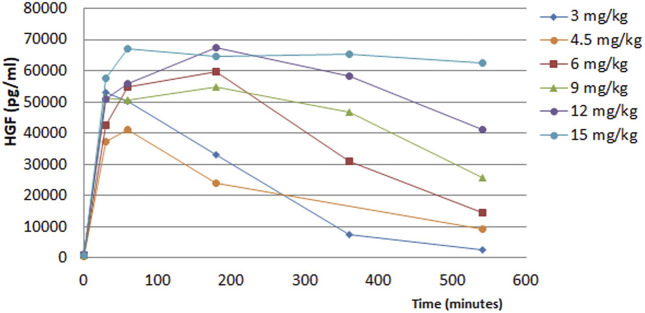
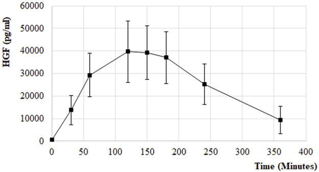
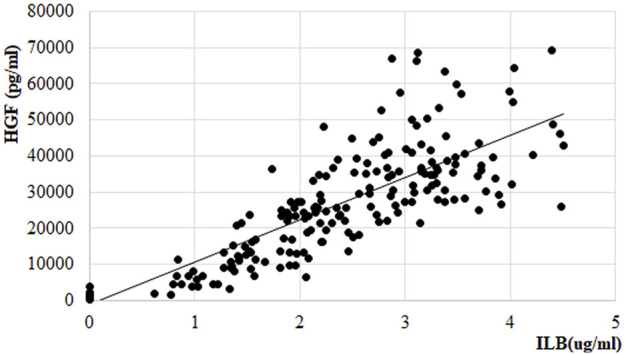
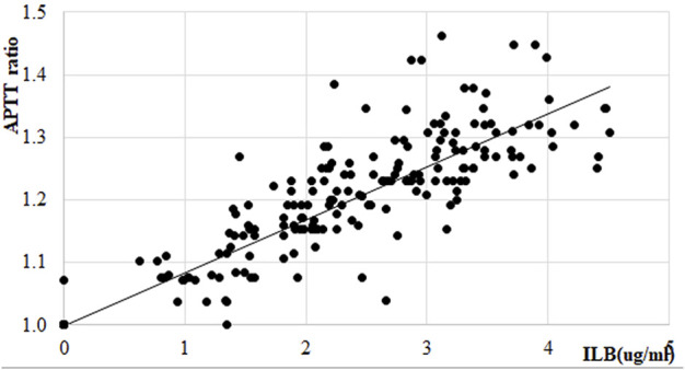
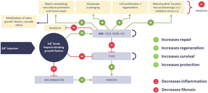
Background: Acute and chronic neurodegenerative diseases represent an immense socioeconomic burden that drives the need for new disease modifying drugs. Common pathogenic mechanisms in these diseases are evident, suggesting that a platform neuroprotective therapy may offer effective treatments. Here we present evidence for the mode of pharmacological action of a novel neuroprotective low molecular weight dextran sulphate drug called ILB®. The working hypothesis was that ILB® acts via the activation of heparin-binding growth factors (HBGF). Methods: Pre-clinical and clinical (healthy people and patients with ALS) in vitro and in vivo studies evaluated the mode of action of ILB®. In vitro binding studies, functional assays and gene expression analyses were followed by the assessment of the drug effects in an animal model of severe traumatic brain injury (sTBI) using gene expression studies followed by functional analysis. Clinical data, to assess the hypothesized mode of action, are also presented from early phase clinical trials. Results: ILB® lengthened APTT time, acted as a competitive inhibitor for HGF-Glypican-3 binding, effected pulse release of heparin-binding growth factors (HBGF) into the circulation and modulated growth factor signaling pathways. Gene expression analysis demonstrated substantial similarities in the functional dysregulation induced by sTBI and various human neurodegenerative conditions and supported a cascading effect of ILB® on growth factor activation, followed by gene expression changes with profound beneficial effect on molecular and cellular functions affected by these diseases. The transcriptional signature of ILB® relevant to cell survival, inflammation, glutamate signaling, metabolism and synaptogenesis, are consistent with the activation of neuroprotective growth factors as was the ability of ILB® to elevate circulating levels of HGF in animal models and humans. Conclusion: ILB® releases, redistributes and modulates the bioactivity of HBGF that target disease compromised nervous tissues to initiate a cascade of transcriptional, metabolic and immunological effects that control glutamate toxicity, normalize tissue bioenergetics, and resolve inflammation to improve tissue function. This unique mechanism of action mobilizes and modulates naturally occurring tissue repair mechanisms to restore cellular homeostasis and function. The identified pharmacological impact of ILB® supports the potential to treat various acute and chronic neurodegenerative disease, including sTBI and ALS.
Keywords: amyotrophic lateral sclerosis; glutamate; heparin-binding growth factors; inflammation; low molecular weight-dextran sulphate; metabolism; neurodegeneration; traumatic brain injury.
Copyright © 2022 Logan, Belli, Di Pietro, Tavazzi, Lazzarino, Mangione, Lazzarino, Morano, Qureshi, Bruce, Barnes and Nagy.
Conflict of interest statement
The authors declare a potential conflict of interest and state it below. Patents pertaining to this LMW-DS drug have been filed by Tikomed AB. LB is coinventor of LMW-DS, and is a founder, shareholder and board member of Tikomed AB. AL, ZN, NB, and LB declare consultancy payments from Tikomed AB and/or Axolotl Consulting Ltd. for services related to the submitted work. IM and OQ are employees of Celentyx Ltd. OQ and NB are shareholders in Celentyx Ltd. NB is a Director of Celentyx Ltd. The remaining authors declare that the research was conducted in the absence of any commercial or financial relationships that could be construed as a potential conflict of interest.
Figures
Similar articles
-
A low molecular weight dextran sulphate, ILB®, for the treatment of amyotrophic lateral sclerosis (ALS): An open-label, single-arm, single-centre, phase II trial.PLoS One. 2024 Jul 11;19(7):e0291285. doi: 10.1371/journal.pone.0291285. eCollection 2024. PLoS One. 2024. PMID: 38990927 Free PMC article. Clinical Trial.
-
ILB®, a Low Molecular Weight Dextran Sulphate, Restores Glutamate Homeostasis, Amino Acid Metabolism and Neurocognitive Functions in a Rat Model of Severe Traumatic Brain Injury.Int J Mol Sci. 2022 Jul 30;23(15):8460. doi: 10.3390/ijms23158460. Int J Mol Sci. 2022. PMID: 35955592 Free PMC article.
-
A phase II open label clinical study of the safety, tolerability and efficacy of ILB® for Amyotrophic Lateral Sclerosis.PLoS One. 2022 May 25;17(5):e0267183. doi: 10.1371/journal.pone.0267183. eCollection 2022. PLoS One. 2022. PMID: 35613082 Free PMC article. Clinical Trial.
-
Glutamate, excitotoxicity and amyotrophic lateral sclerosis.J Neurol. 1997 May;244 Suppl 2:S3-14. doi: 10.1007/BF03160574. J Neurol. 1997. PMID: 9178165 Review.
-
HGF and MET: From Brain Development to Neurological Disorders.Front Cell Dev Biol. 2021 Jun 9;9:683609. doi: 10.3389/fcell.2021.683609. eCollection 2021. Front Cell Dev Biol. 2021. PMID: 34179015 Free PMC article. Review.
Cited by
-
Circular RNA expression in ALS is progressively deregulated and tissue-dependent.BMC Genomics. 2025 Jul 1;26(1):576. doi: 10.1186/s12864-025-11725-4. BMC Genomics. 2025. PMID: 40596818 Free PMC article.
-
A low molecular weight dextran sulphate, ILB®, for the treatment of amyotrophic lateral sclerosis (ALS): An open-label, single-arm, single-centre, phase II trial.PLoS One. 2024 Jul 11;19(7):e0291285. doi: 10.1371/journal.pone.0291285. eCollection 2024. PLoS One. 2024. PMID: 38990927 Free PMC article. Clinical Trial.
References
LinkOut - more resources
Full Text Sources
Other Literature Sources
Miscellaneous