. 2022 Aug 30:13:975676.
doi: 10.3389/fimmu.2022.975676. eCollection 2022.

An optimized Factor H-Fc fusion protein against multidrug-resistant Neisseria gonorrhoeae

Affiliations

An optimized Factor H-Fc fusion protein against multidrug-resistant Neisseria gonorrhoeae

Jutamas Shaughnessy et al. Front Immunol. .

Abstract

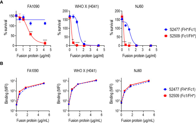
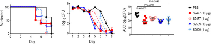
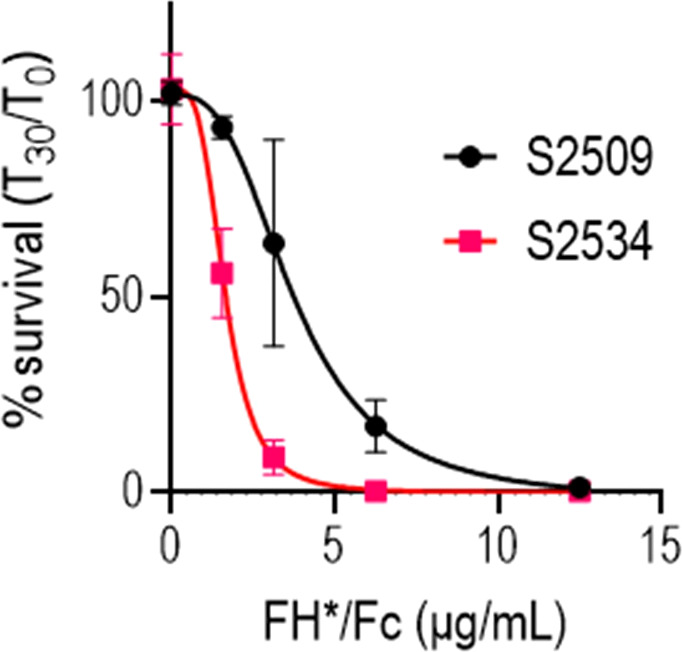
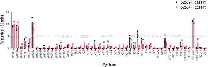
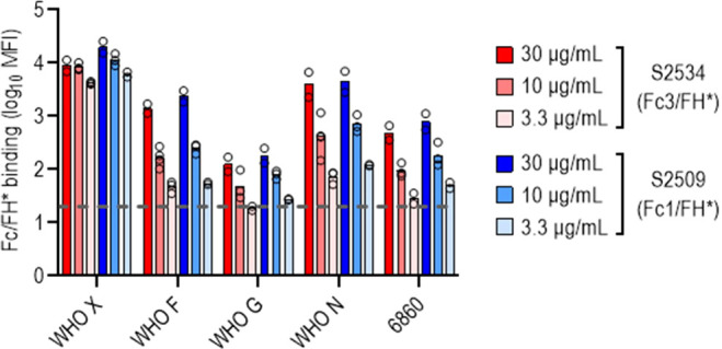
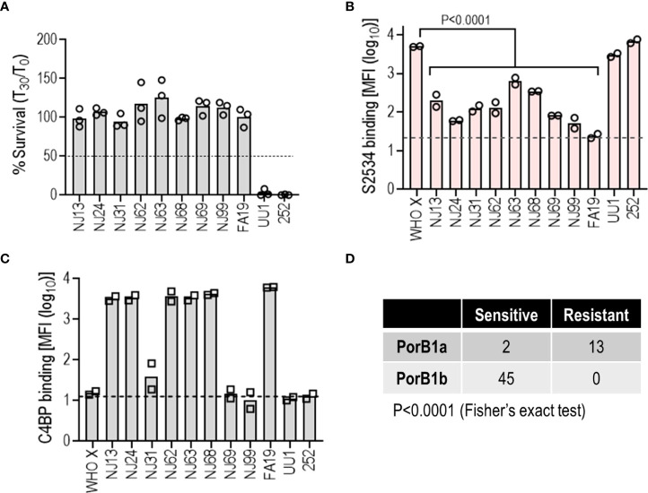
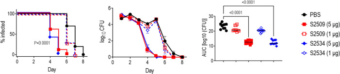
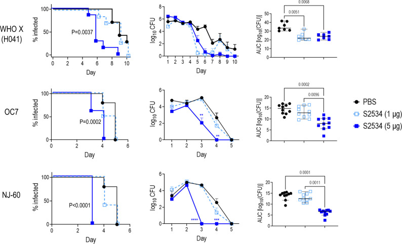
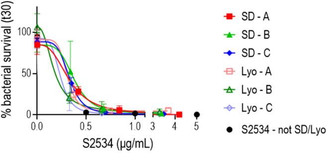
Novel therapeutics against the global threat of multidrug-resistant Neisseria gonorrhoeae are urgently needed. Gonococci evade killing by complement by binding factor H (FH), a key inhibitor of the alternative pathway. FH comprises 20 short consensus repeat (SCR) domains organized as a single chain. Gonococci bind FH through domains 6 and 7, and C-terminal domains 18 through 20. Previously, we showed that a chimeric protein comprising (from the N- to C-terminus) FH domains 18-20 (containing a point mutation in domain 19 to prevent lysis of host cells) fused to human IgG1 Fc (called FH*/Fc1) killed gonococci in a complement-dependent manner and reduced the duration and bacterial burden in the mouse vaginal colonization model of gonorrhea. Considering the N. gonorrhoeae-binding FH domains 18-20 are C-terminal in native FH, we reasoned that positioning Fc N-terminal to FH* (Fc1/FH*) would improve binding and bactericidal activity. Although both molecules bound gonococci similarly, Fc1/FH* displayed a 5-fold lower IC50 (the concentration required for 50% killing in complement-dependent bactericidal assays) than FH*/Fc1. To further increase complement activation, we replaced human IgG1 Fc in Fc1/FH* with Fc from human IgG3, the most potent complement-activating IgG subclass, to obtain Fc3/FH*. Bactericidal activity was further increased ~2.3-fold in Fc3/FH* compared to Fc1/FH*. Fc3/FH* killed (defined by <50% survival) 45/45 (100%) diverse PorB1B-expessing gonococci, but only 2/15 PorB1A-expressing isolates, in a complement-dependent manner. Decreased Fc3/FH* binding accounted for the limited activity against PorB1A strains. Fc3/FH* was efficacious against all four tested PorB1B gonococcal strains in the mouse vaginal colonization model when administered at a dose of 5 µg intravaginally, daily. Furthermore, Fc3/FH* retained bactericidal activity when reconstituted following lyophilization or spray-drying, suggesting feasibility for formulation into intravaginal rings. In conclusion, Fc3/FH* represents a promising prophylactic immunotherapeutic against multidrug-resistant gonococci.

Keywords: Fc fusion protein; Neisseria gonorrhoeae; Nicotiana benthamiana; complement; factor H; gonorrhea; immunotherapeutic.


Conflict of interest statement

Authors YT, JM, and KW were employed by Planet Biotechnology, Inc. The remaining authors declare that the research was conducted in the absence of any commercial or financial relationships that could be construed as a potential conflict of interest.

Figures

Figure 1
Figure 1
Production, characterization and stability of S2477 (FH*/Fc1), S2509 (Fc1/FH*) and S2534 (Fc3/FH*). (A). SDS-PAGE visualized with StainFree (Left image) and western blots probed with anti-human IgG (Right image) of Protein A purified S2477 (FH*/Fc1) (lanes 1 and 6), S2509 (Fc1/FH*) (lanes 2 & 7), and S2534 (Fc3/FH*) (lanes 3 and 8) under non-reducing (lanes 1-3) and reducing conditions (lanes 6-8). (B). Yields of the fusion proteins. The Table shows that yields of S2477 (FH*/Fc1) and S2534 (Fc3/FH*) purified by Protein A chromatography were similar over multiple processing runs. Only one processing run was carried out for S2509 (Fc1/FH*) but the observed yield was within the ranges observed for S2477 and S2534. (C) Stability of S2509 and S2477. Left graph: S2509 (Fc1/FH*) was more stable than S2477 (FH*/Fc1) over 76 days at 37°C. Right graph: Replacing human IgG1 Fc (S2509) with IgG3 Fc (S2534) improves the stability of Fc/FH* in three different buffers over 74 days at 40°C. VFS, simulated vaginal fluid that contains 77 mM NaCl, 7 mM lactic acid, 13 mM acetic acid, 25 mM glucose, 4 mM sodium acetate, 36 mM sodium lactate, pH 6. HRE (Histidine Arginine Glutamic acid buffer) contains 50 mM histidine, 36 mM arginine, 57 mM glutamic acid, 75 mM NaCl, pH 7.
Figure 2
Figure 2
Bactericidal activity and binding of S2477 (FH*/Fc1) and S2509 (Fc1/FH*) produced in N. benthamiana against N. gonorrhoeae in vitro. (A) Serum bactericidal activity of S2477 (FH*/Fc1) and S2509 (Fc1/FH*). The proteins were incubated (concentrations indicated in the X-axis) with N. gonorrhoeae strains FA1090, WHO X (H041) and NJ60 and 20% human complement. Survival of bacteria following incubation for 30 min relative to CFU at 0 min is expressed as a percentage on the Y-axis. The mean (SEM) of 4 separate experiments is shown. The best-fit curve used to generate the IC50 is shown. Two-way ANOVA was used to measure differences in killing across the two molecules at each concentration. **P<0.01; ***P<0.001; ****P<0.0001. (B) Binding of S2477 (FH*/Fc1) and S2509 (Fc1/FH*) to N. gonorrhoeae. The fusion proteins (concentrations indicated on the X-axis) to N. gonorrhoeae FA1090, H041 and NJ60 was measured by flow cytometry. The median fluorescence intensity (MFI) is indicated on the Y-axis. The mean (range) of two separate experiments is shown.
Figure 3
Figure 3
Efficacy of S2477 (FH*/Fc1) and S2509 (Fc1/FH*) against N. gonorrhoeae FA1090 in human FH/C4BP transgenic mice. Premarin®-treated 6-8 week-old human FH/C4BP transgenic mice (n=8/group) were infected with 2.8 x 10⁷ CFU N. gonorrhoeae FA1090. Mice were treated daily (starting 2 h before infection) intravaginally either with PBS (vehicle control) or with 1 µg or 10 µg of S2477 or S2509. Left graph: Kaplan Meier curves showing time to clearance, analyzed the Mantel-Cox (log-rank) test. Middle graph: log10 CFU versus time. X-axis, day; Y-axis, log10 CFU. Comparisons of the CFU over time between each treatment group and the respective saline control was made by two-way ANOVA and Dunnett’s multiple comparison test. All treatment groups showed statistically significantly lower CFUs (P<0.01) compared to the PBS group on day 5 and (with the exception of S2509, 1 µg/d) day 6. Right graph: Bacterial burdens consolidated over time (Area Under the Curve [log10 CFU] analysis). The AUC for each mouse was derived from the log10 CFU data shown in the middle graph. The five groups were compared by one-way ANOVA using the non-parametric Kruskal-Wallis equality of populations rank test. The χ2 with ties 22.77 (P = 0.0001). Pairwise AUC comparisons across groups was made with Dunn’s multiple comparison test.
Figure 4
Figure 4
Replacing human IgG1 Fc with human IgG3 Fc improves bactericidal activity of FH*/Fc. N. gonorrhoeae FA1090 was incubated with increasing concentrations (indicated on the X-axis) of S2509 (Fc1/FH*) or S2534 (Fc3/FH*) and human complement (IgG/IgM depleted human serum) and survival at 30 min was measured (Y-axis). The mean (range) of 3 experiments is shown.
Figure 5
Figure 5
Complement-dependent bactericidal activity of S2509 (Fc1/FH*) and S2534 (Fc3/FH*) against a panel of 50 strains of N. gonorrhoeae. Fifty strains of N. gonorrhoeae (listed on the X-axis; see Supplemental Table S1 for a description of strains) were incubated with the fusion proteins (33 µg/mL) and human complement (IgG and IgM depleted human serum; final concentration 10%). Bacterial survival at 30 min relative to 0 min is indicated as a percentage on the Y-axis. The mean (range) of two separate experiments is shown. Complement alone did not result in any killing of any strain (survival >100%) (data not shown).
Figure 6
Figure 6
N. gonorrhoeae resistant to S2534 (Fc3/FH*) bind lower amounts of the fusion proteins. Binding of S2509 (Fc1/FH*) and S2534 (Fc3/FH*) to the four strains of N. gonorrhoeae that were resistant to killing by S2509 (>50% survival in Figure 5 ) was measured by flow cytometry. WHO X (H041) that was fully susceptible to S2509 and S2534 (100% killing) was included as a comparator. The Y-axis shows the log10 median fluorescence intensity (MFI) of fusion protein binding (mean and individual values of 2-4 separate experiments is shown). The dashed grey line indicates the average value of the conjugate controls (21; range from 11-37).
Figure 7
Figure 7
Low binding of S2534 (Fc3/FH*) limits its efficacy against PorB1A N. gonorrhoeae. (A) Complement-dependent bactericidal activity of S2534 (Fc3/FH*) against PorB1A gonococcal isolates. Ten additional PorB1A-expressing strains of N. gonorrhoeae (data with strain 252, used as a positive control for killing, has been shown in Figure 6 ) were incubated with S2534 (33 µg/mL) and human complement (IgG and IgM depleted human serum; final concentration 10%). Bacterial survival at 30 min relative to 0 min is indicated as a percentage on the Y-axis. The mean (range) of two separate experiments is shown. Complement alone did not result in any killing of any strain (survival >100%) (data not shown). (B) Binding of S2534 (Fc3/FH*; 3.3 µg/mL) to 11 PorB1A-expressing N. gonorrhoeae was measured by flow cytometry. WHO X (PorB1B) that was fully susceptible to S2534 (100% killing) was included as a comparator. The Y-axis shows the log10 median fluorescence intensity (MFI) of fusion protein binding (mean and individual values of 2 separate experiments). The dashed grey line indicates the average value of the conjugate controls (25.7). Comparisons across groups was made with one-way ANOVA and pairwise comparisons with WHO X were made by Dunnett’s multiple comparison test. (C) Binding of C4BP to N. gonorrhoeae. The 11 PorB1A N. gonorrhoeae strains shown in panel B and PorB1B strain WHO X were incubated with 10% normal human serum (NHS) as a source of C4BP. Bound C4BP was detected with mAb 104. Binding is expressed as log10 median fluorescence intensity (MFI) on the Y-axis (mean and individual values of two separate experiments). The dashed grey line indicates the average value of the conjugate controls (14.2). (D) The efficacy of S2534 correlates with the PorB molecule expressed. Bactericidal data from Figures 6 , 7A were compiled into the Table and analyzed with Fisher’s exact test.
Figure 8
Figure 8
Efficacy of S2509 (Fc1/FH*) and S2534 (Fc3/FH*) against N. gonorrhoeae FA1090 in human FH/C4BP transgenic mice. Estrogen pellet-treated 6 week-old human FH/C4BP transgenic mice (n=10/group) were infected with 2.4 x 10⁷ CFU N. gonorrhoeae strain FA1090. Mice were treated daily (starting 2 h before infection) intravaginally either with PBS (vehicle control) or with 1 or 5 µg of each fusion protein. Left graph: Kaplan Meier curves showing time to clearance, analyzed the Mantel-Cox (log-rank) test. Both treatment groups that received 5 µg/d showed significantly faster clearance (P < 0.0001) compared to each of the other groups when compared in a pairwise manner. Middle graph: log10 CFU versus time. X-axis, day; Y-axis, log10 CFU. Comparisons of the CFU over time between each treatment group and the respective saline control was made by two-way ANOVA and Dunnett’s multiple comparison test. The two groups that received 5 µg of the fusion protein daily showed significantly lower CFUs compared to the PBS groups on days 4, 5 and 6 (P < 0.0001, P < 0.0001 and P < 0.05, respectively). Right graph: Bacterial burdens consolidated over time (Area Under the Curve [log10 CFU] analysis). The five groups were compared by one-way ANOVA using the non-parametric Kruskal-Wallis equality of populations rank test. The χ2 with ties was 39.11 (P < 0.0001). Pairwise AUC comparisons across groups was made with Dunn’s multiple comparison test.
Figure 9
Figure 9
Efficacy of S2534 (Fc3/FH*) against three strains of N. gonorrhoeae. Premarin®-treated 6 week-old human FH/C4BP transgenic mice were infected with strains WHO X (H041) (top row; inoculum 2.5 x 10⁷ CFU; n = 7 mice/group), OC7 (middle row; inoculum 3.8 x 10⁷ CFU; n = 10 mice/group) or NJ60 (bottom row; inoculum 3.2 x 10⁷ CFU; n = 10 mice/group) and treated daily (starting 2 h before infection) intravaginally either with PBS (vehicle control) or with 1 or 5 µg of S2534. Left graphs: Kaplan Meier curves showing time to clearance, analyzed the Mantel-Cox (log-rank) test. P values indicate comparisons between the PBS and 5 µg groups. Middle graphs: log10 CFU versus time. X-axis, day; Y-axis, log10 CFU. Comparisons of the CFU over time between each treatment group and the respective saline control was made by two-way ANOVA. Right graph: Bacterial burdens consolidated over time (Area Under the Curve [log10 CFU] analysis). The three groups were compared by one-way ANOVA using the non-parametric Kruskal-Wallis equality of populations rank test. The χ2 with ties were 11.78 (P = 0.0007), 17.11 (P = 0.0002) and 19.61 (P < 0.0001) for WHO X (H041), OC7 and NJ60, respectively. Pairwise AUC comparisons across groups was made with Dunn’s multiple comparison test and the significant P values are indicated.
Figure 10
Figure 10
S2534 (Fc3/FH*) retains bactericidal activity after spray drying or lyophilizing. S2534 was spray-dried (SD) or lyophilized (Lyo) using three different formulations (A, B or C), as shown in Table 2 . The bactericidal activity of each formulation reconstituted in water at concentrations of S2534 ranging from 0 to 5 µg/mL against strain FA1090 was determined. Each data point shows the mean (standard error) of 3 separate experiments. The calculated IC50 for SD – A, SD – B, SD – C, Lyo – A, Lyo – B and Lyo – C were 0.32, 0.38, 0.32, 0.29, 0.17 and 0.22 µg/mL, respectively. The IC50 for the native (not SD or lyophilized) S2534 could not be calculated because 97% killing was seen at the lowest concentration (0.5 µg/mL) tested. There were no statistical differences across the SD or Lyo groups by two-way ANOVA.

References

    1. Rowley J, Vander Hoorn S, Korenromp E, Low N, Unemo M, Abu-Raddad LJ, et al. . Chlamydia, gonorrhoea, trichomoniasis and syphilis: global prevalence and incidence estimates, 2016. Bull World Health Organ (2019) 97:548–562P. doi: 10.2471/BLT.18.228486 - DOI - PMC - PubMed
    1. Laga M, Manoka A, Kivuvu M, Malele B, Tuliza M, Nzila N, et al. . Non-ulcerative sexually transmitted diseases as risk factors for HIV- 1 transmission in women: results from a cohort study [see comments]. Aids (1993) 7:95–102. doi: 10.1097/00002030-199301000-00015 - DOI - PubMed
    1. Cohen CR, Sinei S, Reilly M, Bukusi E, Eschenbach D, Holmes KK, et al. . Effect of human immunodeficiency virus type 1 infection upon acute salpingitis: a laparoscopic study. J Infect Dis (1998) 178:1352–8. doi: 10.1086/314465 - DOI - PubMed
    1. Cohen MS, Hoffman IF, Royce RA, Kazembe P, Dyer JR, Daly CC, et al. . Reduction of concentration of HIV-1 in semen after treatment of urethritis: implications for prevention of sexual transmission of HIV-1. AIDSCAP Malawi research group. Lancet (1997) 349:1868–73. doi: 10.1016/S0140-6736(97)02190-9 - DOI - PubMed
    1. Unemo M, Seifert HS, Hook EW, 3rd, Hawkes S, Ndowa F, Dillon. JR. Gonorrhoea. Nat Rev Dis Primers (2019) 5:79. doi: 10.1038/s41572-019-0128-6 - DOI - PubMed

Publication types