. 2022 Aug 30:12:939166.
doi: 10.3389/fonc.2022.939166. eCollection 2022.

Chondroitin sulfate proteoglycan 4 expression in chondrosarcoma: A potential target for antibody-based immunotherapy

Affiliations

Chondroitin sulfate proteoglycan 4 expression in chondrosarcoma: A potential target for antibody-based immunotherapy

Sjoerd P F T Nota et al. Front Oncol. .

Abstract

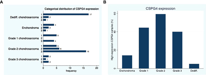
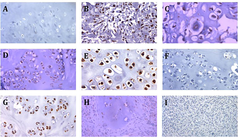
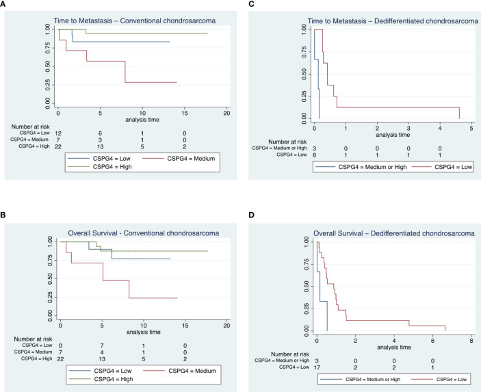
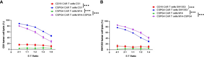
Chondrosarcoma is a common primary bone malignancy whose phenotype increases with its histologic grade. They are relatively resistant to chemotherapy and radiation therapy limiting curative options for disseminated disease. Chondroitin sulfate proteoglycan 4 (CSPG4) is a cell surface proteoglycan that is highly expressed across various human cancers, including chondrosarcoma, and has restricted distribution in healthy tissues, making it an attractive target for the antibody-based therapy. CSPG4 specific chimeric antigen receptor (CAR) T cell therapies have been shown to be effective in treating other cancers such as melanoma and triple negative breast cancer. The goal of this study was to assess the prevalence of CSPG4 in human chondrosarcoma and to assess the efficacy of CSPG4 specific CAR T cells in lysing chondrosarcoma cells in vitro. Using immunohistochemistry (IHC), we stained a tissue microarray containing primary conventional and dedifferentiated chondrosarcoma from 76 patients with CSPG4 specific monoclonal antibodies (mAbs). In addition, we incubated 2 chondrosarcoma cell lines with CSPG4-targeting CAR T cells and subsequently evaluated cell survival. Our results showed medium to high expression of CSPG4 in 29 of 41 (71%) conventional chondrosarcoma tumors and in 3 of 20 (15%) dedifferentiated chondrosarcoma tumors. CSPG4 expression showed a positive association with time to metastasis and survival in both subtypes. CSPG4 CAR T treated cell lines showed a lysis of respectively >80% and 70% demonstrating CSPG4-targeted CAR T cells effective in killing CSPG4-positive chondrosarcoma tumors.

Keywords: CAR T; CSPG4; chondrosarcoma; dedifferentiated chondrosarcoma; immunotherapy.


Conflict of interest statement

The authors declare that the research was conducted in the absence of any commercial or financial relationships that could be construed as a potential conflict of interest.

Figures

Figure 1
Figure 1
Distribution of CSPG4 Expression in Chondrosarcoma. (A) CSPG4 staining using IHC was scored by 2 investigators using a categorical classification, 1-low expression; 2-medium expression; 3 high-expression. (B) The number of samples within each category that stained positive with high intensity (categorical classification 3) was plotted as a percentage of the samples in the cohort; the distribution of high intensity CSPG4 positive samples showed a unimodal distribution with the highest percentage observed in grade II. Fisher’s exact p = 0.001.
Figure 2
Figure 2
Representative images of CSPG4 staining of primary chondrosarcoma tumors. (A) CSPG4 negative stain in enchondroma (200x magnification). (B) CSPG4 positive stain in grade 2 chondrosarcoma (200x magnification). (C) CSPG4 positive stain in grade 2 chondrosarcoma (400x magnification). (D) CSPG4 positive stain in grade 2 chondrosarcoma (200x magnification). (E) CSPG4 positive stain in grade 2 chondrosarcoma (400x magnification). (F) CSPG4 negative stain in grade 2 chondrosarcoma (200x magnification). (G) CSPG4 positive stain in grade 2 chondrosarcoma (200x magnification). (H) CSPG4 positive stain in dedifferentiated chondrosarcoma (200x magnification). (I) CSPG4 negative stain in dedifferentiated chondrosarcoma (200x magnification).
Figure 3
Figure 3
Time to metastasis and overall survival in conventional and dedifferentiated chondrosarcoma. (A, B) All patients diagnosed with chondrosarcoma grade 1 (n = 17) remained alive throughout the duration of the study, 7 out of the 30 subjects diagnosed with chondrosarcoma grade 2 and 2 out of 5 patients with chondrosarcoma grade 3 died from their disease. We observed a shorter time to metastasis in subjects with medium CSPG4 expression compared to the other groups in CSPG4 expression (p = 0.004) and there was a shorter time to death in subjects with a medium CSPG4 expression (p = 0.019). (C, D) All 24 patients with a dedifferentiated chondrosarcoma died from their disease. We observed a shorter time to metastasis in subjects who had medium and high CSPG4 expression when compared to subjects with low or no CSPG4 expression in chondrosarcoma (p = 0.048). In addition overall survival was shorter in subject who exhibited medium and high CSPG4 expression compared to subjects with low or no CSPG4 expression in chondrosarcoma tissues (p = 0.024).
Figure 4
Figure 4
CSPG4-CART cells are effective in killing CSPG4-expressing chondrosarcoma cells. CSPG4 CAR T cells and target cells were co-cultured at indicated (E) T. ratios for 24 hrs. CAR T cells in the cell suspension were removed, and the viability of adherent target cells was quantitated by MTT assays. Mean ± SEM of cell lysis (%) of different cell populations in the chondrosarcoma cell line CS1 (A) and SW1353 (B) are shown. The human melanoma cell line Ml4 which does not express CSPG4 and M14/CSPG4 which express CSPG4 after stably transfected with CSPG4 plasmid DNA were used as specificity controls25. CD19 CAR T cells were also used as a negative control since none of the target cells express CD19. The experiments were performed in triplicate and repeated 3 times. Differences between CSPG4 CART cell-mediated cytotoxicity on different cell populations (including all E .T ratios) was detected using a chi square test in a two-way ANOVA. ***p < 0.001.

References

    1. Limaiem F, Davis DD, Sticco KL. Chondrosarcoma. StatPearls. Treasure Island (FL: StatPearls Publishing Copyright © 2022, StatPearls Publishing LLC. (2022).
    1. Shariat Torbaghan S, Ashouri M, Jalayer Naderi N, Baherini N. Histopathologic differentiation between enchondroma and well-differentiated chondrosarcoma: Evaluating the efficacy of diagnostic histologic structures. J Dental Res Dental Clin Dental Prospects (2011) 5:98–101. doi: 10.5681/joddd.2011.022 - DOI - PMC - PubMed
    1. Strotman PK, Reif TJ, Kliethermes SA, Sandhu JK, Nystrom LM. Dedifferentiated chondrosarcoma: A survival analysis of 159 cases from the SEER database (2001-2011). J Surg Oncol (2017) 116:252–7. doi: 10.1002/jso.24650 - DOI - PubMed
    1. Stevenson JD, Laitinen MK, Parry MC, Sumathi V, Grimer RJ, Jeys LM. The role of surgical margins in chondrosarcoma. Eur J Surg Oncol J Eur Soc Surg Oncol Br Assoc Surg Oncol (2018) 44:1412–8. doi: 10.1016/j.ejso.2018.05.033 - DOI - PubMed
    1. Italiano A, Mir O, Cioffi A, Palmerini E, Piperno-Neumann S, Perrin C, et al. . Advanced chondrosarcomas: role of chemotherapy and survival. Ann Oncol (2013) 24:2916–22. doi: 10.1093/annonc/mdt374 - DOI - PMC - PubMed

LinkOut - more resources