. 2022 Sep 7:21:331-341.
doi: 10.1016/j.reth.2022.08.005. eCollection 2022 Dec.

Personalized tissue-engineered arteries as vascular graft transplants: A safety study in sheep

Affiliations

Personalized tissue-engineered arteries as vascular graft transplants: A safety study in sheep

Lachmi Jenndahl et al. Regen Ther. .

Abstract

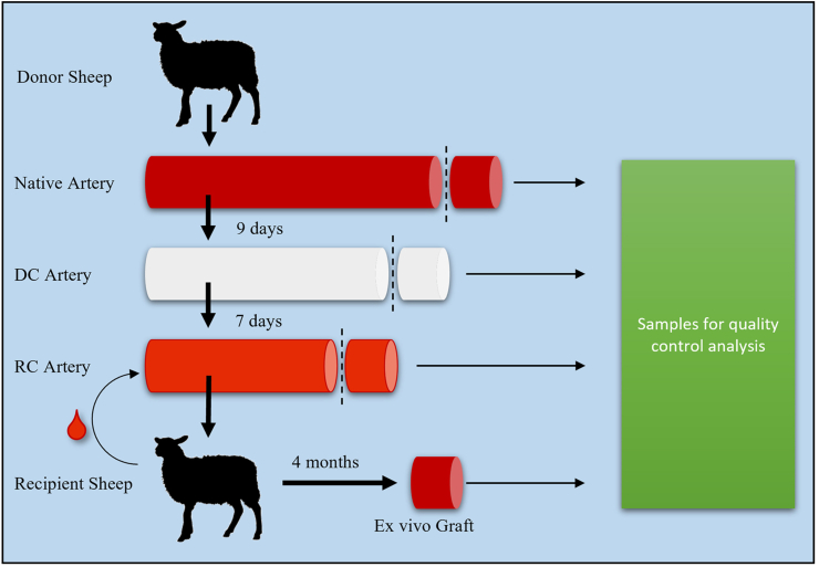
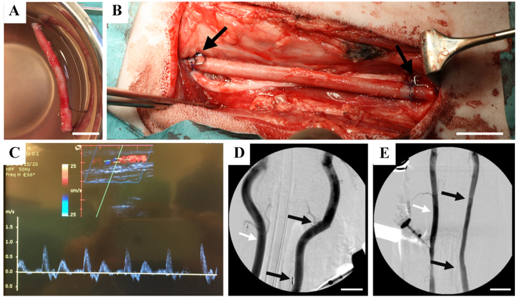
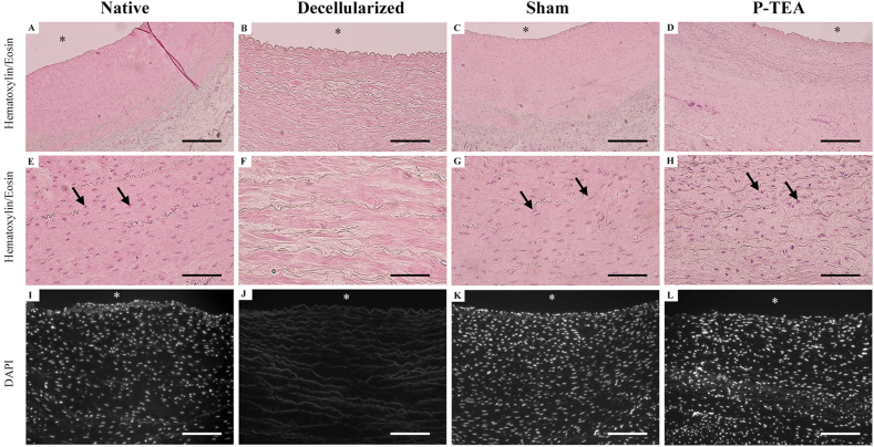
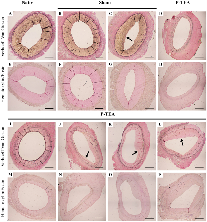
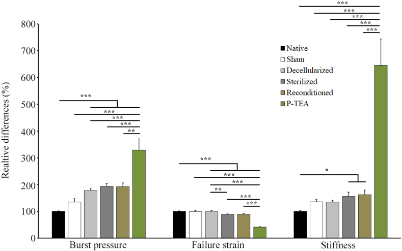
Patients with cardiovascular disease often need replacement or bypass of a diseased blood vessel. With disadvantages of both autologous blood vessels and synthetic grafts, tissue engineering is emerging as a promising alternative of advanced therapy medicinal products for individualized blood vessels. By reconditioning of a decellularized blood vessel with the recipient's own peripheral blood, we have been able to prevent rejection without using immunosuppressants and prime grafts for efficient recellularization in vivo. Recently, decellularized veins reconditioned with autologous peripheral blood were shown to be safe and functional in a porcine in vivo study as a potential alternative for vein grafting. In this study, personalized tissue engineered arteries (P-TEA) were developed using the same methodology and evaluated for safety in a sheep in vivo model of carotid artery transplantation. Five personalized arteries were transplanted to carotid arteries and analyzed for safety and patency as well as with histology after four months in vivo. All grafts were fully patent without any occlusion or stenosis. The tissue was well cellularized with a continuous endothelial cell layer covering the luminal surface, revascularized adventitia with capillaries and no sign of rejection or infection. In summary, the results indicate that P-TEA is safe to use and has potential as clinical grafts.

Keywords: ATMP; Blood vessels; Recellularization; Regenerative medicine; Scaffold; Tissue engineering.


Conflict of interest statement

Lachmi Jenndahl, Tobias Gustafsson-Hedberg, Robin Simsa and Raimund Strehl were employees of the company VERIGRAFT AB which contributed financially, including salaries and study costs, and by providing laboratory space.

Figures

Fig. 1
Fig. 1
Schematic overview of the graft through experimental process. Carotid arteries are donated from sheep slaughter waste, decellularized over 9 days, reconditioned with the recipient sheep’s peripheral blood over seven days and the personalized tissue engineered artery is transplanted to the carotid artery. Four months after surgery, the graft is excised. Samples for quality control are harvested in all steps. DC = Decellularization, RC = Reconditioning.
Fig. 2
Fig. 2
Transplantation of carotid artery on sheep and patent blood vessel four months after transplantation. (A) Reconditioned personalized tissue engineered artery (P-TEA) before transplantation. (B) Transplantation of the P-TEA to the carotid artery of sheep with end-to-end anastomosis. (C) Representative ultrasound image of P-TEA four months after surgery showing the patent artery with the graph illustrating adequate blood flow. (D, E) shows angiography from sham and P-TEV, respectively, with patent blood vessels without stenosis or occlusion. Black arrows in B, D and E marks metal clips sutured on the outside of the anastomosis for localization of the surgical site during angiography. Left vessel in D and E (marked with white arrow), shows the native non-operated artery at the other side of trachea. Scale bars are 2 cm.
Fig. 3
Fig. 3
Hematoxylin/Eosin and 4′,6-diamidino-2-phenylindole (DAPI) stainings of carotid artery after four months in vivo. The cells in the native arteries (A, E, I) were efficiently removed in the decellularization process (B, F, J). After four months in vivo, sham operated arteries (C, G, K) and the P-TEAs (D, H, L) were comparable cellularized with native artery (A, E, I). Arrows in E, G and H indicate cell nuclei. No nuclei were identified in B, F or J. ∗ = luminal side. Scale bars are 200 μm in A– D and I – L; 50 μm in E − H.
Fig. 4
Fig. 4
Characterization of the blood vessel tissue after four months in vivo. The luminal surface was lined with cells expressing the endothelial cell marker CD31 (red), and cell nuclei are indicated with DAPI (blue) (A-D). All cells in the native tissue (A) were removed during the decellularization (B). The sham operated arteries (C) and the personalized tissue engineered arteries (P-TEA) (D) were equivalent with the native tissue four months after transplantation. Scanning electron microscopy shows the endothelial cells on the luminal surface of native artery (E) which were efficiently removed after decellularization (F). Reconditioning added a layer of blood components and cells (G), and the luminal surface was completely recellularized after four months in vivo (H). Staining with antibodies against CD31 (red) and alpha smooth muscle actin (αSMA) (green) as well as SYTOX™ Deep Red Nucleic Acid Stain (blue) for nuclei staining during P-TEA production process (I–N). The αSMA expression in native carotid artery cells (I) remained in the tissue after the decellularization (J) and reconditioning (K) process. After four months in vivo, the cells in P-TEA showed intracellular expression of αSMA (L) in a remodeled fashion compared with native tissue. Arrows marks cell nuclei and intracellular expression of αSMA. Confocal images of the adventitia in native carotid artery (M) and P-TEA (N) shows capillary revascularization with CD31 expressing cells lined with αSMA-expressing smooth muscle cells and/or pericytes after four months in vivo. Von Kossa staining shows no trace of calcification of the P-TEA after four months in vivo as comparing native (O) and P-TEA (P). Insets are larger magnifications of the same image. ∗ = luminal side. Scale bars are 200 μm in A – D, I – L and O, P; 150 μm in M, N and 50 μm in inset I and L.
Fig. 5
Fig. 5
Characterization of intimal hyperplasia in tissue engineered arteries after four months in vivo. Hematoxylin/eosin and Van Gieson staining’s illustrates the extracellular matrix and intimal hyperplasia (A–P). Native carotid artery without any initial hyperplasia (A, E). Sham operated carotid artery four months after surgery with sections of areas with no (B, F) and small (C, G) intimal hyperplasia. Personalized tissue engineered arteries after four months in vivo showing areas with no (D, H, I, M) small (J, N) some (K, O) or more (L, P) intimal hyperplasia. Arrows indicate intimal hyperplasia. Scale bars are 100 μm.
Fig. 6
Fig. 6
Biomechanical properties of artery during the tissue engineering process and after four months in vivo. Samples from individual arteries of native (n = 36), sham operated (n = 2), decellularized (n = 36), sterilized (n = 36), reconditioned (n = 36) and after four months in vivo (n = 5) were stretched until failure with a biomechanical testing device and analyzed for burst pressure, failure strain and stiffness. ∗ = p < 0.05, ∗∗ = p < 0.01, ∗∗∗ = p < 0.001.

Similar articles

Cited by

References

    1. Blann A.D. How a damaged blood vessel wall contributes to thrombosis and hypertension. Pathophysiol Haemostasis Thromb. 2003;33(5–6):445–448. doi: 10.1159/000083843. - DOI - PubMed
    1. Chemla E.S., Morsy M. Randomized clinical trial comparing decellularized bovine ureter with expanded polytetrafluoroethylene for vascular access. Br J Surg. 2009;96(1):34–39. doi: 10.1002/bjs.6434. - DOI - PubMed
    1. Cines D.B., Pollak E.S., Buck C.A., Loscalzo J., Zimmerman G.A., McEver R.P., et al. Endothelial cells in physiology and in the pathophysiology of vascular disorders. Blood. 1998;91(10):3527–3561. - PubMed
    1. Conte M.S., Bandyk D.F., Clowes A.W., Moneta G.L., Seely L., Lorenz T.J., et al. Results of PREVENT III: a multicenter, randomized trial of edifoligide for the prevention of vein graft failure in lower extremity bypass surgery. J Vasc Surg. 2006;43(4):742–751. doi: 10.1016/j.jvs.2005.12.058. discussion 751. - DOI - PubMed
    1. Crapo P.M., Gilbert T.W., Badylak S.F. An overview of tissue and whole organ decellularization processes. Biomaterials. 2011;32(12):3233–3243. doi: 10.1016/j.biomaterials.2011.01.057. - DOI - PMC - PubMed

LinkOut - more resources