. 2022 Aug;10(16):852.
doi: 10.21037/atm-22-1105.

A comprehensive framework of the right posterior section for tailored anatomical liver resection based on three-dimensional simulation system

Affiliations

A comprehensive framework of the right posterior section for tailored anatomical liver resection based on three-dimensional simulation system

Feihong Zhang et al. Ann Transl Med. 2022 Aug.

Abstract

Background: The anatomical right posterior sectionectomy (ARPS) is a technically challenging procedure. We aimed to develop and validate a novel framework of the right posterior section for a safe and tailored anatomical liver resection (ALR) based on a three-dimensional (3D) simulation system.

Methods: 3D hepatectomy simulations of healthy participants who underwent contrast-enhanced computed tomography of the upper abdomen were retrospectively reviewed to develop the framework according to the relationship between the simulated plane determined by the right posterior portal pedicle (RPP) and the course of the right hepatic vein (RHV) trunk. The framework was validated in the practice of ARPS for hepatocellular carcinoma (HCC) prospectively.

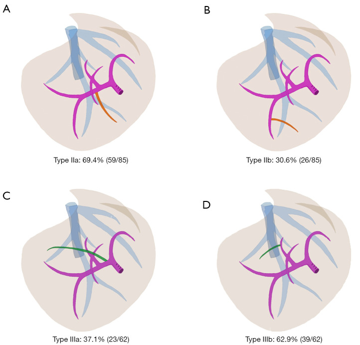
Results: Scans from 336 eligible participants were assessed. The framework was summarized into four types: normal, caudal-redundant, cranial-deficient, and combined types, accounting for 43.4% (146/336), 25.3% (85/336), 18.5% (62/336), and 12.8% (43/336) respectively. The caudal-redundant type was associated with the variable portal branches of the RPP or segment 6 branch across the ventral side of RHV. The mean aberrant volume proportion in type IIa was significantly greater than that in type IIb (P<0.001), which were 7.0%±3.5% and 4.4%±1.8% respectively. The cranial-deficient type was associated with the aberrant segment 7 portal pedicle originating from the right portal trunk or the dorsal portal branch of segment 8 crossing over to the RHV. The median aberrant volume proportion in type IIIa was significantly greater than that in type IIIb (P<0.001), which were 10.9% (8.5-13.3%) and 4.0% (3.0-6.1%), respectively. The combined type represented a combination of the caudal-redundant type and the cranial-deficient type. The framework provided instructions on tailored ARPS in 6 patients with HCC by maximizing lesion removal and functional liver remnant with favorable perioperative outcomes.

Conclusions: Precise preoperative planning with an individualized surgical approach based on our framework allows safe anatomical liver resections for cases with lesions in the right posterior section.

Keywords: Anatomical liver resection (ALR); computed tomography; outcomes; right posterior section; three-dimensional visualization.


Conflict of interest statement

Conflicts of Interest: All authors have completed the ICMJE uniform disclosure form (available at https://atm.amegroups.com/article/view/10.21037/atm-22-1105/coif). The authors have no conflicts of interest to declare.

Figures

Figure 1
Figure 1
Flowchart of study design. CECT, contrast-enhanced computed tomography; DICOM, Digital Imaging and Communications in Medicine; 3D, three-dimensional.
Figure 2
Figure 2
Morphological framework of the right posterior section. (A) Normal type (type I): the intersegmental border determined by the RPPT coincides with the course of the RHV trunk; (B) Caudal-redundant type (type II): the intersegmental border determined by the RPPT protrudes caudal-laterally against the course of the RHV trunk; (C) Cranial-deficient type (type III): the intersegmental border determined by the RPPT sinks cranial-laterally against the course of the RHV trunk; (D) Combined type (type IV): Type II + III. Note that the white dotted line indicates the course of the RHV trunk. The pink region represents the RPP-oriented territory, and the blue region represents the future liver remnant. RPPT, right posterior portal territory; RPP, right posterior portal pedicle; RHV, right hepatic vein.
Figure 3
Figure 3
Schematic diagram of portal pedicle variations in the framework. (A) Type IIa: caudal-redundant type with Ventral-P6 crossing over to the RHV; (B) type IIb: caudal-redundant type with the variant distal portal branch of P6; (C) type IIIa: cranial-deficient type with RP-7; (D) type IIIb: cranial-deficient type with P8c crossing over to the RHV. Note that aberrant portal branches of type II and type III are highlighted in orange and green. P6, portal pedicle of segment 6; Ventral-P6, the aberrant P6 running across the ventral side of RHV; RP-7, the aberrant portal branch of segment 7 originated from the right portal vein; P8c, dorsal portal branch of segment 8; RHV, right hepatic vein.
Figure 4
Figure 4
Schematic diagram of portal pedicle variations in type IV. (A) Type IVa: type IIa combined with type IIIa; (B) type IVb: type IIa combined with type IIIb; (C) type IVc: type IIb combined with type IIIa; (D) type IVd: type IIb combined with IIIb. Aberrant portal branches of type II and type III are highlighted in orange and green.
Figure 5
Figure 5
Tailored surgical strategies according to the framework of the right posterior section. Note that the white dotted line indicates the demarcation plane oriented by the RPPT and the red dotted line represents the course of the RHV trunk. RPPT, right posterior portal territory; RHV, right hepatic vein; ARPS, anatomical right posterior sectionectomy; S8d, dorsal subsegment 8; Ventral-P6, the aberrant P6 running across the ventral side of RHV.
Figure 6
Figure 6
Tailored laparoscopic ARPS combined with S8d resection for recurrent hepatocellular carcinoma in a patient with type IVb variant. (A) Preoperative CECT revealed the tumor in segment 7 (orange bold arrow). (B,C) Preoperative 3D simulation showed type IVb variant. The RPPT (pink region) failed to cover the whole tumor between segment 7 and segment 8. Aberrant Ventral-P6s (type IIa variation) that one originated near the hepatic hilum (white arrow) and the other located far away from the hepatic hilum (black arrow), combined with P8c (yellow arrow) crossing over to the RHV (type IIIb variation), were noted. Tailored transection plane of parenchyma-sparing ARPS combined with S8d resection (black dotted line) was simulated after preserving the first Ventral-P6 that originates near the hepatic hilum (white arrow). (D) The root of RPP was exposed by blunt dissection after preserving the first Ventral-P6 (white arrow) at the level of Rouviere’s sulcus. The second ventral-P6 was dissected for fear of postoperative ischemia. (E,F) Intraoperative ICG stain was identical to preoperative 3D simulation. Negative counterstaining of partial segment 6 fed by the second Ventral-P6 allowed for precise liver parenchyma transection before mobilizing the right liver. Note that the white dotted line indicates the course of the RHV trunk. ARPS, anatomical right posterior sectionectomy; S8d, dorsal subsegment 8; CECT, contrast-enhanced computed tomography; 3D, three-dimensional; type IV: type IIa combined with type IIIb; RPPT, right posterior portal territory; Ventral-P6, the aberrant portal pedicle of segment 6 running across the ventral side of RHV; P8c, dorsal portal branch of segment 8; RHV, right hepatic vein; RPP, right posterior portal pedicle; ICG, indocyanine green.
Video S1
Video S1
RHV-oriented laparoscopic parenchyma-sparing anatomical right posterior sectionectomy combined with S8d resection.

Comment in

References

    1. Shindoh J, Makuuchi M, Matsuyama Y, et al. Complete removal of the tumor-bearing portal territory decreases local tumor recurrence and improves disease-specific survival of patients with hepatocellular carcinoma. J Hepatol 2016;64:594-600. 10.1016/j.jhep.2015.10.015 - DOI - PubMed
    1. Margonis GA, Buettner S, Andreatos N, et al. Anatomical Resections Improve Disease-free Survival in Patients With KRAS-mutated Colorectal Liver Metastases. Ann Surg 2017;266:641-9. 10.1097/SLA.0000000000002367 - DOI - PubMed
    1. Liao KX, Chen L, Ma L, et al. Laparoscopic middle-hepatic-vein-guided anatomical hemihepatectomy in the treatment of hepatolithiasis: a 10-year case study. Surg Endosc 2022;36:881-8. 10.1007/s00464-021-08344-x - DOI - PubMed
    1. Jang JS, Cho JY, Ahn S, et al. Comparative Performance of the Complexity Classification and the Conventional Major/Minor Classification for Predicting the Difficulty of Liver Resection for Hepatocellular Carcinoma. Ann Surg 2018;267:18-23. 10.1097/SLA.0000000000002292 - DOI - PubMed
    1. Kawaguchi Y, Fuks D, Kokudo N, et al. Difficulty of Laparoscopic Liver Resection: Proposal for a New Classification. Ann Surg 2018;267:13-7. 10.1097/SLA.0000000000002176 - DOI - PubMed