. 2022 Aug 30:10:932983.
doi: 10.3389/fcell.2022.932983. eCollection 2022.

A heterozygous p.S143P mutation in LMNA associates with proteasome dysfunction and enhanced autophagy-mediated degradation of mutant lamins A and C

Affiliations

A heterozygous p.S143P mutation in LMNA associates with proteasome dysfunction and enhanced autophagy-mediated degradation of mutant lamins A and C

Gun West et al. Front Cell Dev Biol. .

Abstract

Lamins A and C are nuclear intermediate filament proteins that form a proteinaceous meshwork called lamina beneath the inner nuclear membrane. Mutations in the LMNA gene encoding lamins A and C cause a heterogenous group of inherited degenerative diseases known as laminopathies. Previous studies have revealed altered cell signaling pathways in lamin-mutant patient cells, but little is known about the fate of mutant lamins A and C within the cells. Here, we analyzed the turnover of lamins A and C in cells derived from a dilated cardiomyopathy patient with a heterozygous p.S143P mutation in LMNA. We found that transcriptional activation and mRNA levels of LMNA are increased in the primary patient fibroblasts, but the protein levels of lamins A and C remain equal in control and patient cells because of a meticulous interplay between autophagy and the ubiquitin-proteasome system (UPS). Both endogenous and ectopic expression of p.S143P lamins A and C cause significantly reduced activity of UPS and an accumulation of K48-ubiquitin chains in the nucleus. Furthermore, K48-ubiquitinated lamins A and C are degraded by compensatory enhanced autophagy, as shown by increased autophagosome formation and binding of lamins A and C to microtubule-associated protein 1A/1B-light chain 3. Finally, chaperone 4-PBA augmented protein degradation by restoring UPS activity as well as autophagy in the patient cells. In summary, our results suggest that the p.S143P-mutant lamins A and C have overloading and deleterious effects on protein degradation machinery and pharmacological interventions with compounds enhancing protein degradation may be beneficial for cell homeostasis.

Keywords: autophagy; degradation; disease mutations; lamin A/C (LMNA); ubiquitin (Ub); ubiquitin-proteasome degradation system.


Conflict of interest statement

The authors declare that the research was conducted in the absence of any commercial or financial relationships that could be construed as a potential conflict of interest.

Figures

FIGURE 1
FIGURE 1
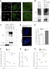
Expression and degradation of lamins A and C are enhanced in LMNA mutant patient cells. (A) RT-qPCR analysis of LMNA expression in the patient fibroblasts carrying a heterozygous p.S143P LMNA mutation. Allele-specific primers detecting either wild-type (c.427T) or mutant (c.427C) alleles, or primers detecting both (LMNA) were used. N = 3 individual experiments. (B) LC-ESI-MS/MS analysis of peptide fragments covering both wild-type and mutant lamins A and C in the patient cells. The sequences specific to the p.143S and p.143P lamin A and C peptide fragments are shown on the top, and the corresponding peaks in the mass spectra are encircled. (C) RT-qPCR analysis of relative overall LMNA expression in the control and patient fibroblasts (N = 3). (D) Luciferase assay measuring activity of an upstream −1.3 kb LMNA promotor sequence in the control and patient fibroblasts (N = 3). (E) Western blot analysis shows protein levels of lamins A and C (LA/C) in the patient and control fibroblasts treated with 300 μg/ml cycloheximide (CHX) for given time points. Numerical values show levels of lamins A and C normalized to actin, which was used as a loading control. (F) Confocal microscopy images from control and patient cells stained for lamins A and C before or after 16-h treatment with 50 µM leptomycin B (LMB). Mid-plane confocal sections are shown. Scale bar 10 µm. (G) Mean fluorescence intensities (AU) were measured from confocal images and plotted (N = 300). Note upregulation of lamins A and C after 16-h treatment with 50 µM LMB in the patient fibroblasts. The whiskers show the mean values ±s.d. and boxplots show the 75th and 25th percentiles of the calculated intensities, *p < 0.05, **p < 0.01, ***p < 0.001.
FIGURE 2
FIGURE 2
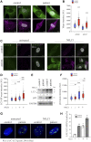
Lamins A and C are ubiquitinated by K48-linked chains in LMNA mutant patient fibroblasts. (A) Representative confocal microscopy images from control and patient fibroblasts stained for lamins A and C and K48-linked ubiquitin chains. Scale bar 10 µm. (B) Pulldown with GST-tagged recombinant pan-tandem ubiquitin-binding entity (pan-TUBE) under denatured conditions. (C) Pulldown with lamin A antibody under denatured conditions. K48-ubiquitin levels were normalized to GAPDH, which was used as a loading control. (D-E) Proximity ligation assay (PLA) of lamins A and C and K48-ubiquitin in the control and patient fibroblasts as calculated from >300 individual cells. Cells with more than three PLA signals were considered positive. Data are expressed as mean ± s.e.m, *p < 0.05. (F) Proteasome activity of control and patient fibroblasts treated with or without 20 mM NH4Cl for 24 h. N = 3 individual experiments. (G) Chymotrypsin-like activity of 20S proteasomes isolated from control and patient fibroblasts (N = 3). (H) Chymotrypsin-like activity of 20S proteasomes isolated from Hela cells expressing either FLAG-tagged WT-LA or p.S143P-LA and treated with or without 20 mM NH4Cl for 24 h (N = 3). The whiskers show mean values ±s.d **p < 0.01, ***p < 0.001.
FIGURE 3
FIGURE 3
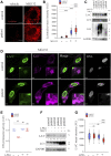
Lamins A and C are increasingly committed to autophagy in LMNA mutant patient fibroblasts. (A) Confocal microscopy images from the patient and control fibroblasts stained for autophagy-related proteins Atg5 and Atg7. Scale bar 10 µm. (B) Calculated mean fluorescence intensity values from Atg5 and Atg7 stainings. The whiskers show mean values ± s.d and boxplots show the 75th and 25th percentiles (N = 300), *** p < 0.001. (C) Confocal microscopy images from the patient and control fibroblasts treated with or without 20 mM NH4Cl for 24h and stained for Atg8/LC3-I/II and SQSTM1/p62. Note the accumulation of LC3-I/II or p62 after NH4Cl treatment especially in the patient cells. Scale bar 10 µm. (D) Calculated mean fluorescence intensity values from LC3-I/II staining. The whiskers show mean values ± s.d and boxplots show the 75th and 25th percentiles (N = 300), *** p < 0.001 (compared to untreated control cells). (E) Western blot analysis from control and patient fibroblasts treated with 20 mM NH4Cl for 24 h. Pooled LC3-I/II and p62 levels were normalized to GAPDH, which was used as a loading control. Note the increase of LC3-II after NH4Cl treatment. (F) Calculated mean fluorescence intensity values from p62 staining. (N = 300), *** p < 0.001 (G–H) Proximity ligation assay (PLA) detecting association of lamins A and C with LC3-I/II in the control and patient fibroblasts. The percentage of cells with PLA signals was determined from >300 cells and the cells with more than three PLA signals were considered positive. Data is expressed as mean ± s.e.m, * p < 0.05, ** p < 0.01, *** p < 0.001.
FIGURE 4
FIGURE 4
Crosstalk between autophagy and UPS is impaired in LMNA mutant patient cells. (A) LysoTracker Red DND-99 staining from untreated and MG132-treated control and patient cells. (B) Calculated mean fluorescence intensity values show more LysoTracker positive lysosomes in the patient cells and their number is further increased after 24-h treatment with 1 µM MG132. (C) Western blot analysis from control and patient cells treated with 1 µM MG132 or 20 mM NH4Cl for 24 h. Protein levels of lamins A and C (LA/C) were normalized to GAPDH, which was used as a loading control. All the proteins were detected on the same membrane. (D) Confocal microscopy images from control and patient cells treated with MG132 and stained for lamins A and C and K48-ubiquitin. Insets show cytosolic structures that co-stain with lamins A and C and K48 antibodies. Scale bar 10 µm. (E) Chymotrypsin-like activity as measured in control and patient cells treated with 5 mM 4-PBA for 24 h (N = 3 individual experiments) **p < 0.01. (F) Western blot analysis of control and patient cells treated with or without 5 mM 4-PBA. Autophagy was inhibited with 20 mM NH4Cl and GAPDH was used as a loading control. (G) Calculated mean fluorescence intensity values (AU) from control and patient fibroblasts treated with or without 5 mM 4-PBA for 24 h and stained for lamins A and C (N = 300). The whiskers show mean values ±s.d and boxplots show the 75th and 25th percentiles, *p < 0.05, ***p < 0.001.
FIGURE 5
FIGURE 5
Illustrative picture of degradation of K48-ubiquitinated lamins A and C. In normal cells, wild-type lamins A and C are partially ubiquitinated with K48-linked chains that lead to UPS-mediated degradation of lamins A and C. In the patient cells, lamins A and C are increasingly K48-ubiquitinated, presumably due to increased turnover and production, as well as proteasomal dysfunction. This leads to saturation of UPS and an accumulation of K48-linked ubiquitin chains within the nucleus. Saturation and dysfunction of UPS further lead to compensatory degradation of K48-lamins A and C through autophagy, created with BioRender.com.

References

    1. Albornoz N., Bustamante H., Soza A., Burgos P. (2019). Cellular responses to proteasome inhibition: molecular mechanisms and beyond. Int. J. Mol. Sci. 20 (14), 3379. 10.3390/ijms20143379 - DOI - PMC - PubMed
    1. Amm I., Sommer T., Wolf D. H. (2014). Protein quality control and elimination of protein waste: the role of the ubiquitin-proteasome system. Biochim. Biophys. Acta 1843, 182–196. 10.1016/j.bbamcr.2013.06.031 - DOI - PubMed
    1. Arrasate M., Mitra S., Schweitzer E. S., Segal M. R., Finkbeiner S. (2004). Inclusion body formation reduces levels of mutant huntingtin and the risk of neuronal death. Nature 431, 805–810. 10.1038/nature02998 - DOI - PubMed
    1. Bachmair A., Varshavsky A. (1989). The degradation signal in a short-lived protein. Cell 56, 1019–1032. 10.1016/0092-8674(89)90635-1 - DOI - PubMed
    1. Bence N. F., Sampat R. M., Kopito R. R. (2001). Impairment of the ubiquitin-proteasome system by protein aggregation. Science 292, 1552–1555. 10.1126/science.292.5521.1552 - DOI - PubMed