. 2022 Nov 18;17(22):e202200327.
doi: 10.1002/cmdc.202200327. Epub 2022 Oct 11.

Identification of 2,4-Dinitro-Biphenyl-Based Compounds as MAPEG Inhibitors

Affiliations

Identification of 2,4-Dinitro-Biphenyl-Based Compounds as MAPEG Inhibitors

Simone Di Micco et al. ChemMedChem. .

Abstract

We identified 2,4-dinitro-biphenyl-based compounds as new inhibitors of leukotriene C4 synthase (LTC4 S) and 5-lipoxygenase-activating protein (FLAP), both members of the "Membrane Associated Proteins in Eicosanoid and Glutathione metabolism" (MAPEG) family involved in the biosynthesis of pro-inflammatory eicosanoids. By molecular docking we evaluated the putative binding against the targets of interest, and by applying cell-free and cell-based assays we assessed the inhibition of LTC4 S and FLAP by the small molecules at low micromolar concentrations. The present results integrate the previously observed inhibitory profile of the tested compounds against another MAPEG member, i. e., microsomal prostaglandin E2 synthase (mPGES)-1, suggesting that the 2,4-dinitro-biphenyl scaffold is a suitable molecular platform for a multitargeting approach to modulate pro-inflammatory mediators in inflammation and cancer treatment.

Keywords: FLAP inhibitors; LTC4S inhibitors; anti-inflammatory drugs; anticancer agents; multitargeting approach.


Conflict of interest statement

The authors declare no conflict of interest.

Figures
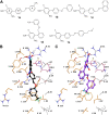
Figure 1
Figure 1
(A) 1 ae chemical structures. Three‐dimensional model of the interactions given by 1 a (B) and 1 d (C) with LTC4S. The protein is represented by orange (chain A) and grey (chain C) tube (C, according to chain colour; polar H, white; N, dark‐blue; O, red; S, yellow), whereas the GSH by faded‐salmon tube with the same atom colour code of the enzyme, except for C (faded‐salmon). The ligands are depicted by sticks (1 a, black; 1 d, violet) and balls (C, as for the sticks; polar H, white; N, dark‐blue; O, red; F, light green). The dashed black and violet lines indicate the H‐bonds and salt bridges, respectively.
Figure 2
Figure 2
Three‐dimensional model of the interactions given by 1 a (A), 1 c (B), 1 d (C) and 1 e (D) with FLAP. The protein is depicted by faded‐yellow (chain A) and green (chain C) tube (C, according to chain colour; polar H, white; N, dark‐blue; O, red). The ligands are represented by sticks (1 a, black; 1 c, cyan; 1 d, violet; 1 e, orange) and balls (C, as for the sticks; polar H, white; N, dark‐blue; O, red; F, light green). The dashed black and violet lines indicate the H‐bonds and salt bridges, respectively.
Figure 3
Figure 3
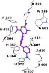
Three‐dimensional model of interactions given by 1 d with 5‐LOX. The protein is depicted by grey tube (C, grey; polar H, white; N, dark‐blue; O, red). The ligand is represented by violet sticks and balls (C, as for the sticks; polar H, white; N, dark‐blue; O, red; F, light green). The dashed black lines indicate the H‐bonds.
Figure 4
Figure 4
Contact histograms during the simulation of 1 d with LTC4S (A), FLAP (B) and 5‐LOX (C).
Figure 5
Figure 5
Catalytic activity of mPGES‐1 in presence of two 1 d concentrations (1 and 10 μM) and after 10‐fold dilution from 10 to 1 μM (=10(1)). The cyan bar indicates the washout salvaged enzymatic activity.
Figure 6
Figure 6
Effect of 1d on 5‐LOX/FLAP interaction in HEK293 cells stably expressing 5‐LOX and FLAP. Cells were pre‐incubated with 1 d (3 μM), MK886 (0.3 μM) or vehicle (0.1 % DMSO) for 10 min at 37 °C and afterwards stimulated by 2.5 μM Ca2+‐ionophore A23187 for 10 min at 37 °C. DAPI (blue) was used to stain the nucleus and proximity ligation assay (PLA) signals (magenta dots) visualize in situ 5‐LOX/FLAP interactions. Results are representative for 100 individual cells analyzed in three independent experiments.

References

    1. Jakobsson P. J., Morgenstern R., Mancini J., Ford-Hutchinson A., Persson B., Am. J. Respir. Crit. Care Med. 2000, 161, S20–S24. - PubMed
    1. Liening S., Scriba G. K., Rummler S., Weinigel C., Kleinschmidt T. K., Haeggstrom J. Z., Werz O., Garscha U., Biochim. Biophys. Acta 2016, 1861, 1605–1613. - PubMed
    1. Yarla N. S., Pathuri G., Gali H., Terzyan S., Panneerselvam J., Chandrakesan P., Scotti M. T., Houchen C., Madka V., Rao C. V., J. Inflammation Res. 2020, 13, 1261–1278. - PMC - PubMed
    1. Thulasingam M., Haeggström J. Z., J. Mol. Biol. 2020, 432, 4999–5022. - PubMed
    1. Werz O., Gerstmeier J., Garscha U., Expert Opin. Ther. Pat. 2017, 27, 607–620. - PubMed

Publication types

MeSH terms

LinkOut - more resources