. 2023 Jan;36(1):78-85.
doi: 10.1111/pcmr.13064. Epub 2022 Oct 9.

Varicella-zoster virus in actively spreading segmental vitiligo skin: Pathological, immunochemical, and ultrastructural findings (a first and preliminary study)

Affiliations

Varicella-zoster virus in actively spreading segmental vitiligo skin: Pathological, immunochemical, and ultrastructural findings (a first and preliminary study)

Yvon Gauthier et al. Pigment Cell Melanoma Res. 2023 Jan.

Abstract

Segmental vitiligo (SV) is a unilateral subtype of vitiligo which is clinically characterized by a cutaneous depigmentation and histologically by a melanocyte loss from the epidermis and hair follicle reservoirs. To date, its pathogenesis remains a mystery. In many cases, this skin depigmentation shares several clinical features and dysfunctions with herpes zoster (HZ). So, for the first time, we examined whether any nucleus and cell fusion associated with a positive immunolabelling of varicella-zoster virus (VZV) and VZV mature virions could be found in SV skin samples as in herpes zoster (HZ). A total of 40 SV samples were used for histological and immunochemical studies. Control samples were obtained from three HZ, and 10 generalized vitiligo lesions. For ultrastructural study, three recent SV and one HZ as controls were recruited. Here, we report that nuclear fusion in epidermal cells were statistically associated with recent SV (p < .001), whereas syncytia formation was associated with long-lasting SV (p = .001). A positive detection of VZV antigen was statistically associated in the epidermis with recent SV and in the dermis with long-lasting SV (p = .001). Finally, the discovery of mature virions in 3/3 recent SV samples provides additional arguments for our viral hypothesis.

Keywords: autonomic nervous system; melanosome transport; nucleus fusion; segmental vitiligo; syncytium; ultrastructural study; varicella-zoster virus; viral cytopathic changes.


Conflict of interest statement

The authors declare that they have no conflict of interest.

Figures
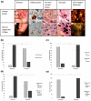
FIGURE 1
FIGURE 1
Histological and immunochemical changes in HZ and in pigmented SV skin. (a) Herpes zoster: Panel a, HZ of left cheek; Panel b, a melanocyte heavily pigmented with large melanin granules in the dendrites (arrow) detected by immunohistochemistry using anti‐HMB45 revealed with DAB in brown, ×40; Panels c and d hematoxylin–eosin‐safran staining showing cell fusion between 2 keratinocytes (arrow), ×40 (c), and a giant polynucleated cell, (arrow) ×40 (d); Panel e, important VZV immunolabelling in epidermal cells (anti VZV antibody revealed using alexa 555 coupled secondary antibody) (white arrows), ×40 IF. Segmental vitiligo: Panel f, recent SV in left cheek; Panel g, marginal melanocytes with several melanin giant granules around the nucleus and within dendrites (arrows), detected by immunohistochemistry using anti‐HMB45 revealed with DAB in brown ×40; Panels h and i Hematoxylin–eosin‐saffron staining showing nuclear fusion between several keratinocytes (black arrows, surrounded in white), ×40 (h) and a large syncytia rarely found in the epidermis (arrow), ×40 (i); Panel J, VZV immunolabeling in an keratinocyte (anti VZV antibody revealed using alexa 555 coupled secondary antibody) (white arrow), ×40. (b) Percentage of SV patients with nuclear fusion (<20 or >20) in epidermis according to the age of SV lesions. (c) Percentage of SV patients without or with syncytia in skin according to the age of the lesions. (d) Percentage of SV patients with negative or positive VZV staining in epidermis according to the age of SV lesions. (e) Percentage of SV patients with negative or positive VZV staining in dermis according to the age of SV lesions. All quantifications were performed by section in recent (n = 13) and long‐lasting vitiligo (n = 27) patients by two blind observers.
FIGURE 2
FIGURE 2
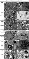
Ultrastructural study of VZV life cycle in HZ and SV and defective melanogenesis in marginal melanocytes of SV. Panel 1: Replication and virus assembly in HZ and SV. (a) VZV capsids (black arrows) in the nucleus, several nucleo capsids (arrowheads) in the nucleus are egressing from the nucleus (N) to perinuclear cisternae, mature virions (white arrow) in the cytoplasm (encart) exit of nucleocapsids through nucleus membrane. (b) several nucleo capsids (black arrows) in nucleus and mature virion (white arrow) in the cytoplasm (encart) nucleocapsids (nc) in the nucleus. Panel 2: Envelopment of nucleocapsids in HZ and SV. (a) (encart) Nucleocapsid (black arrow) adheres to the concave surface of the electron dense sac (S) from Golgi apparatus during the envelopment. (b) envelopment of nucleocapsids (black arrow), (encart) envelopment of nucleo capsid (arrow heads) with electron dense sac (S). Panel 3: Maturation of virions. (a) Numerous nucleocapsids (arrow heads) acquire a characteristic secondary lipid envelopment and become mature virions (white arrows). Encart: a typical VZV mature virion with a nucleo capsid enclosed in a double layer lipid envelope. (b) Several similar mature virions (white arrows) and nucleocapsids (arrow heads) and vesicules (v) in the cytoplasm. Encart: similar aspect of a mature virion. Defective melanogenesis in SV melanocytes infected with VZV. Panel 4: Perinuclear clustering of mature melanosomes and lysosomes. (a) a marginal melanocyte loaded with melanin with a perinuclear clustering of mature melanosomes (black arrow), « honey combed » aspect of the cytoplasm with many large vacuoles (V) Nucleus (N). (b) a perinuclear clustering of large and normal sized melanosomes (black arrow) and lysosomes (L) close to nucleocapsids (arrow heads). Panel 5: Giant melanin granule (a) aggregation of normal and large melanosomes (M), close by a large lysosome (L), encart nucleocapsid (white arrow) in melanin granule. (b) abnormal granule results of the fusion of large and normal sized melanosomes. Panel 6: Lysis of melanosomes. (a) aggregation of several degraded melanosomes (m) close by mature virions (white arrows) and nucleocapsids (arrow heads) (b) a large autophagic vacuole containing residual bodies nearby nucleocapsids (arrow heads) and aggregated melanosomes.

Comment in

Similar articles

Cited by

References

    1. Abdelmaksoud, A. , Vestita, M. , & Lotti, T. (2019). Valacyclovir as a potential therapy for mycosis fungoides and vitiligo in a zosteriform pattern. Dermatologic Therapy, 32, e12870. 10.1111/dth.12870 - DOI - PubMed
    1. Arnozan, X. , & Lenoir, L. (1922). Vitiligo avec troubles nerveux sympathiques: l’ origine sympathique du vitiligo. Bulletin de la Société Française de Dermatologie et de Syphiligraphie, 12, 124–140.
    1. Attili, V. R. , & Attili, S. K. (2013). Segmental and generalized vitiligo: Both forms demonstrate inflammatory histopathological features and clinical mosaicism. Indian Journal of Dermatology, 58, 433–438. - PMC - PubMed
    1. Attili, V. R. , & Attili, S. K. (2015). Anatomical segmentations in cell forms of vitiligo: A new dimension of etiopathogenesis. Indian Journal of Dermatology, Venereology and Leprology, 82(4), 379–388. - PubMed
    1. Barra, D. C. , & Seabra, M. C. (2004). The melanosomes as a model to study organelle motility in mammals. Pigment Cell Research, 17(2), 111–118. - PubMed